In this series of articles I am going to show you some of the exhibits contained in the Museum of Urology, hosted on the BAUS website (www.baus.org.uk).
We are very lucky with the excellent and detailed imaging our radiology colleagues provide for us in the way of ultrasound, CT and MRI. In this issue I am joined by Kamran Raza, one of our core trainees, to tell us about a rather obscure method of renal imaging, which, I imagine, few of you will have even heard of.
Most of us will recall the rude awakening by a pager going off in the early hours of the morning followed by a request to cannulate a patient urgently so that they can attend their IV contrast CT scan. As the on-call junior house officer, you would head to the ward and after a couple of unsuccessful fatigued attempts in a poorly lit bedspace, you finally manage to get a blue cannula into a questionable vein on the patient’s thumb. Satisfied with your efforts, you hurry back to your on-call room to catch a few more minutes of sleep before your next inevitable bleep. Now, spare a thought for Mr William Barr Stirling’s house officer, who instead had the task of cannulating the patient’s abdominal aorta to facilitate an aortogram!
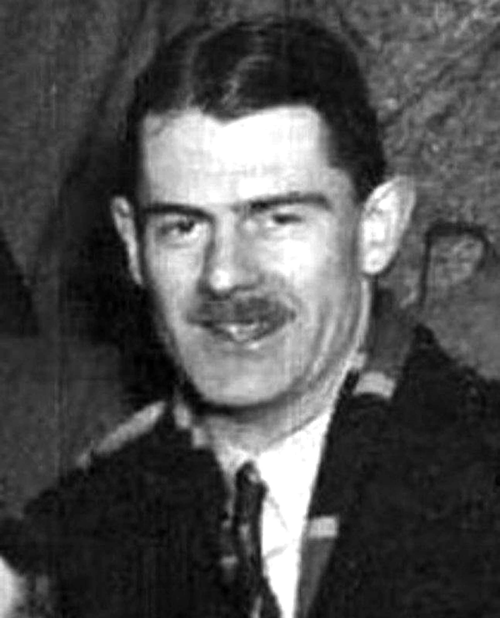
Figure 1: William Barr Stirling, 1936 (reproduced with kind permission of University of
Glasgow Archives & Special Collections, Papers of Angus M Thomson, GB248 DC286/1/76).
William Barr Stirling (1913-1984) was born in Paisley, Scotland on 8 August 1913 (Figure 1). He was enrolled into school at the Glasgow Academy by his father Hugh Stirling, a local tobacco manufacturer, before following in the footsteps of his older brother Hugh by gaining a place at the University of Glasgow to study medicine in 1932. Despite earning accolades as an undergraduate in diseases of the ear, clinical medicine and obstetrics & gynaecology, it was a career in urology, which awaited Barr Stirling. Two years after graduating in 1937 he enrolled into the Royal Navy, serving as a fleet surgeon during World War II. He served as Surgeon Lieutenant on HMS Matabele in the 1940 Norwegian Campaign prior to being transferred to Ceylon to help establish a large naval hospital. Shortly after his transfer, HMS Matabele was torpedoed resulting in the loss of all hands. Both William and Hugh Stirling served for the full duration of the war and remained on the reserve list for a number of years after its conclusion. Posthumously, he was awarded the Arctic Star Medal.
After the war ended, William Barr Stirling married Dr Hannah Stirling MBE (nee Ness), a conservation campaigner, and began work as a urologist in the Glasgow Royal Infirmary. In 1948 he was made a fellow of both the Royal College of Surgeons of Edinburgh and the Royal Faculty of Physicians & Surgeons of Glasgow, and was made Chief of the Urology Department in 1964, succeeding the retired Arthur Jacobs (1899-1974).
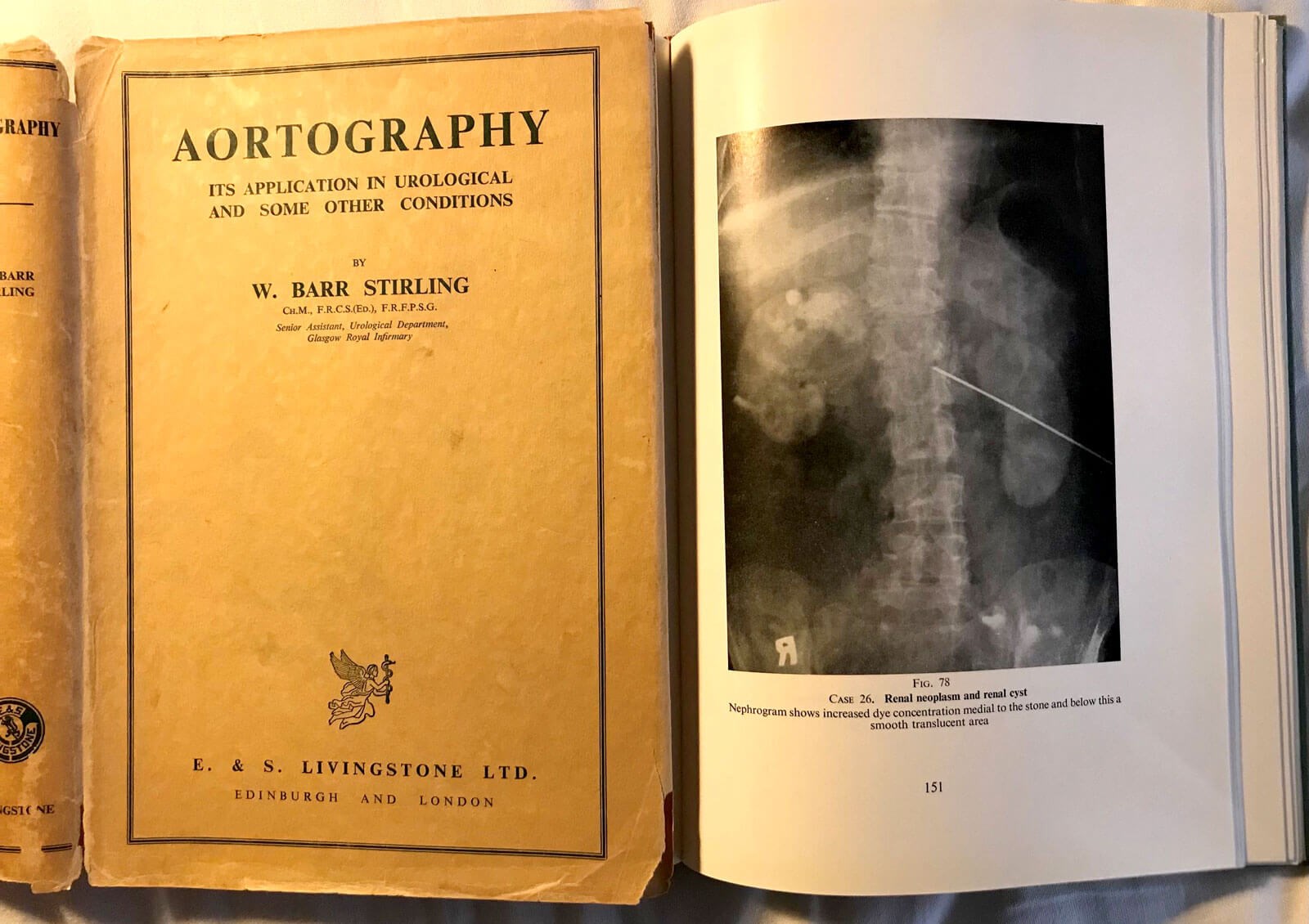
Figure 2: Stirling’s famous book (author’s collection) open showing an aortogram
with the long needle in situ and identifying a right renal tumour.
It was during these years that William Barr Stirling gained published for his book Aortography. Its Application in Urological and Some Other Conditions (Figure 2). Published in 1957, it shares Barr Stirling’s experiences of around 500 aortographs, typically performed for renal disease. The book highlighted the importance of aortography in assessing the function and distribution of blood supply in renal disease and other conditions requiring reparative or conservative surgery. In the kidney, it facilitated diagnosis of congenital abnormalities, cysts, neoplasms, hydronephrosis, calculus, hypoplasia and renal tuberculosis. In his book, Barr Stirling described using a trans-lumbar approach to the aorta and detailed his technique in cannulation of the blood vessel and administration of the contrast medium, as well as use of anaesthesia to carry out the procedure. Essentially, this involved a very long needle and a high-pressure hand pump to inject the contrast at a high rate to allow good opacification of the high flowing aorta and renal vasculature. Considerable skill was needed to get good images.
Aortography was not a new concept; an American study had been conducted on dogs in 1936, but 50% of the test subjects died. This caused fears around the use of aortography in humans, despite evidence showing that the high mortality rate in dogs was due to anatomical differences and a disproportionately higher dose of sodium iodide contrast. Notably, dogs have smaller aortas than humans and when punctured with the same gauge of needle, there is a higher rate of haemorrhage. Nevertheless, whilst there were subsequent cases of aortography in humans, there remained reluctance for its use among the wider surgical community. William Barr Stirling showed that it could be done and his book, described by some as “a classic of its day”, earned him the rightful title as the ‘Pioneer of the aortogram’.
Despite the good renal images the aortogram facilitated, its use in medicine was relatively short-lived. Only 10 years following the publication of William Barr Stirling’s book, the CT scanner was invented by Sir Godfrey Hounsfield (1919-2004) and within five years this was made commercially available. Whilst it took a further two decades before the introduction of CT angiography, much of the pathology which required Barr Stirling’s aortography for diagnosis could now be detected by a simple CT scan, leaving very few indications for the more invasive aortograph.
William Barr Stirling continued working as the Urology Department Chief at Glasgow Royal Infirmary, maintaining a keen interest in academic work and becoming a renowned authority on surgery of the adrenal glands. Aside from his clinical work, William Barr Stirling served as BAUS Treasurer and a council member from 1960 to 1963. He was also elected President of the Section of Urology of the Royal Society of Medicine (1975-1976) and was a member of the International Society of Urologists. He finally retired in 1977 and continued living at his home in Tarbet, Loch Lomond until his sudden death on 1 April 1984. His work in pioneering aortography eventually led to him obtaining ChM (with honours) from the University of Glasgow.
There is little doubt that William Barr Stirling made a significant contribution to urological surgery and was rightfully rewarded with some praise at the time and acclamation at the time. Despite this, is seems that his work was largely overshadowed by the subsequent introduction of CT scanning which left little utility for aortography. The quick succession of CT scanning left no time for aortography to truly leave its mark on surgery and is perhaps the reason why William Barr Stirling’s work never gained the level of historical acclaim for which it was due.