In this series of articles I am going to show you some of the exhibits contained in the Museum of Urology, hosted on the BAUS website (www.baus.org.uk).
I think we all have stories of objects inserted into the urethra and bladder, which we have been obliged to remove. I had naively assumed this was a rather modern practice (you’d think I would have learned not to make that mistake by now).
However, at a recent visit to the EAU headquarters in Arnhem as a member of their History Office, I had the opportunity to examine some of their antique instruments, which made me realise that this is not a new phenomenon. This then prompted me to work with the EAU History Office to arrange a new exhibition in the BAUS Museum of Urology using some of their wonderful collection to explore this area further.
Firstly, we should ask the question, “Why did (and still do) people insert objects into their urethras?” The answer is not necessarily what you may first think. After all, we, as urologists, often insert instruments via the urethra for diagnostic and therapeutic reasons.
In 1783, General Martin, of Lucknow, could stand the pains of his bladder stone no longer and passed a small file into his own bladder several times a day to break it up; it took nine months. Reginald Harrison (1837–1908) working in the port of Liverpool, before he later moved to St Peter’s in London, was commonly faced with chronic gonococcal urethral strictures in sailors returning from long voyages. Harrison described attempts made aboard ship by the ailing sailor or one of the crew to ‘treat’ the stricture by passing a wire or skewer or piece of whittled wood and even on one occasion a piece of lead gas pipe down the urethra. We still see patients now who, in frustration with their urinary symptoms, pass homemade ‘dilators’ into their urethras (in my experience, often retired engineers, and the ‘instruments’ can be very beautifully machined!).
There are iatrogenic foreign bodies. Occasionally, urological instruments break and pieces have to be removed, this occurred in the past as well as now. Fragments of catheters are also relatively ‘frequent flyers’ in this regard.
Older rubber catheters and bougies were more fragile and more likely to fragment. In 1910, Leopold Casper (1859–1959) wrote that broken off catheters and bougies were more likely candidates than self-inserted objects (Figure 1).
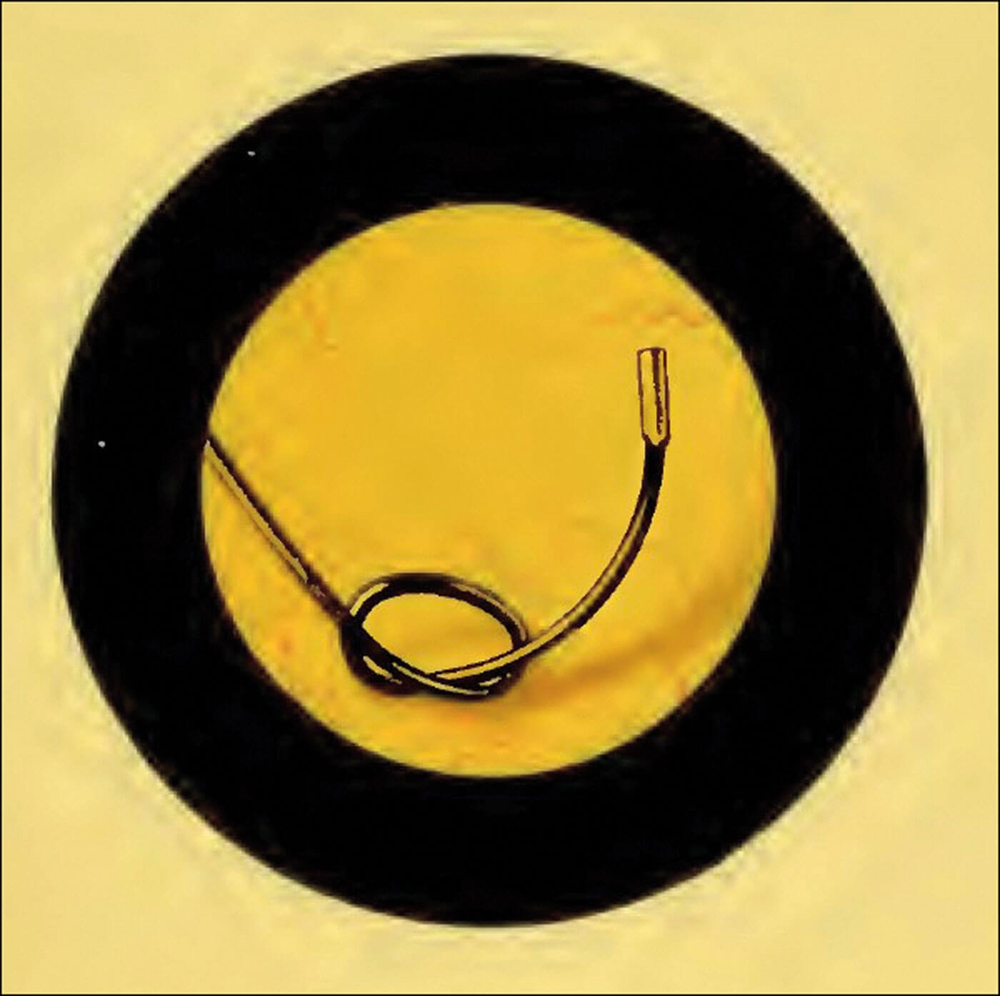
Figure 1: A filiform bougie ‘lost’ in the bladder.
Tinted cystoscopic photograph by Edwin Hurry Fenwick, 1893.
However, quite often the reason was (and is) sexual stimulation, or as Dr Casper called it in 1910, “erotic intensions”. Reginald Harrison took a more sanguine tone saying it was, “for reasons often best known to the patients themselves”. Manipulation of the urethra can, in some, lead to sexual pleasure. The passing of metal or glass rods (often urological dilators) and sometimes fluids, is now popularly known as ‘urethral sounding’, but, as I said, this is not a modern phenomenon.
In 1700, Sir Thomas Molyneux (1661–1733) described a case to The Royal Society of a young Irish Women called Dorcas Blake. She had pushed a bodkin into her bladder, although, the girl swore she had swallowed it. It had to be removed by a very early suprapubic cystotomy. Hairpins, the more modern version of Doras’ bodkin often find their way into female bladders and sometimes remain long enough to act as a nidus for stone formation. According to Emil Zuckerkandl (1849–1910) wax pushed into the bladder does not encrust, silver does so slowly, while iron, rubber and vegetable materials encrust quickly, forming a phosphatic stone.
Hairpins, it appears were quite a popular object to insert. Edward Hurry Fenwick (1856–1944) of the London Hospital took an early photograph of one with a Nitze Photo-Cystoscope. Instruments specifically designed to remove hairpins feature in the Collin’s Instrument Catalogue of 1925. The lists of objects inserted, found and removed from the human bladder, described in old textbooks, are varied and impressive and I’m sure we can all add to this (Figure 2).
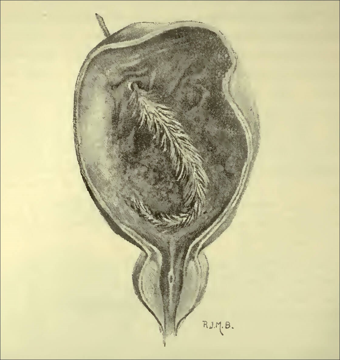
Figure 2: A grass stalk pushed into the urethra and lost by a patient of Reginald Harrison’s
in Liverpool in 1865. A reluctant historian, in an age before cystoscopy, the patient died of peritonitis.
Occasionally the presentation can be obscure, as Thomson Walker said, “the history of the introduction of the foreign body may be hard to elicit” and the urologist may find they are trying to explain a diagnosis of cystitis, haematuria or infection with an incomplete history. Casper felt that he could generally get a clear history, and if not, in that modern age, he possessed “a sovereign means of diagnosis in the cystoscope”. In 1916, Edward Canny Ryall, a pioneer endourologist from All Saints’ Hospital discovered a hairpin in a 41-year-old lady’s bladder; she denied all knowledge of it initially, eventually confessing that she had pushed it up six or eight weeks before.
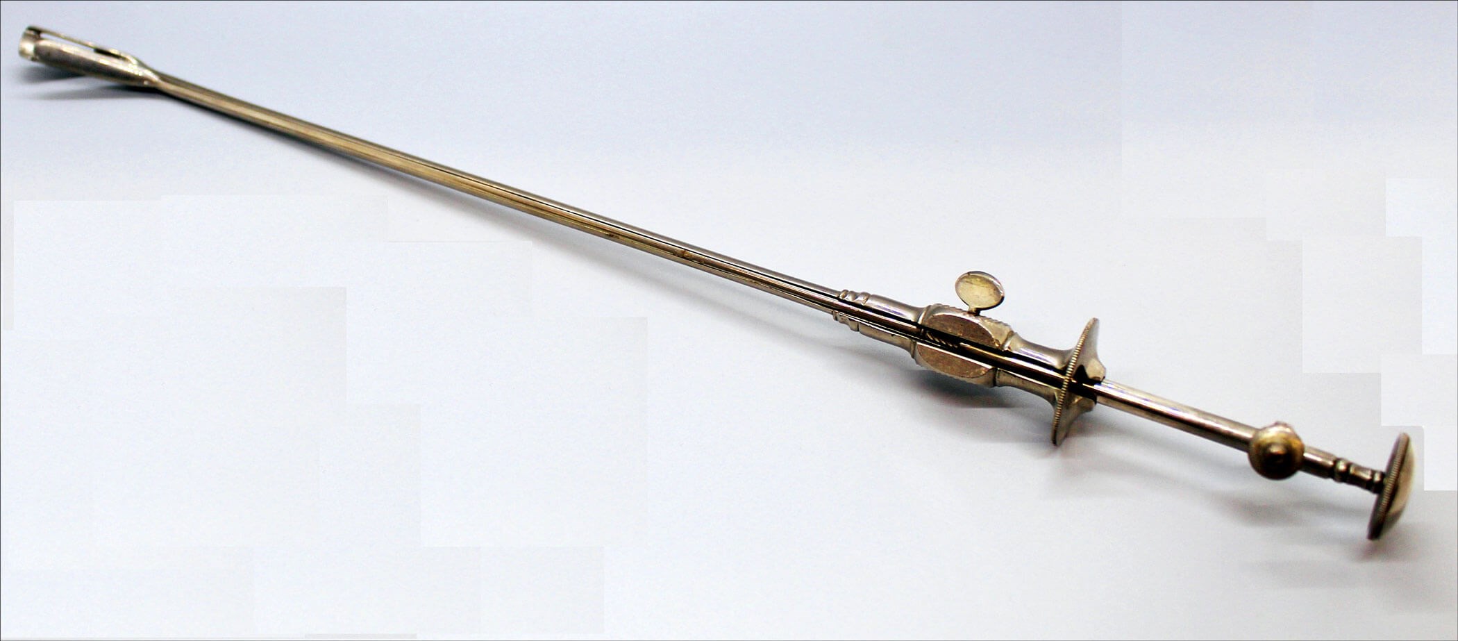
Figure 3a: Instrument for removing foreign bodies from the male bladder, Collin, Paris.
From the EAU History Office Collection.
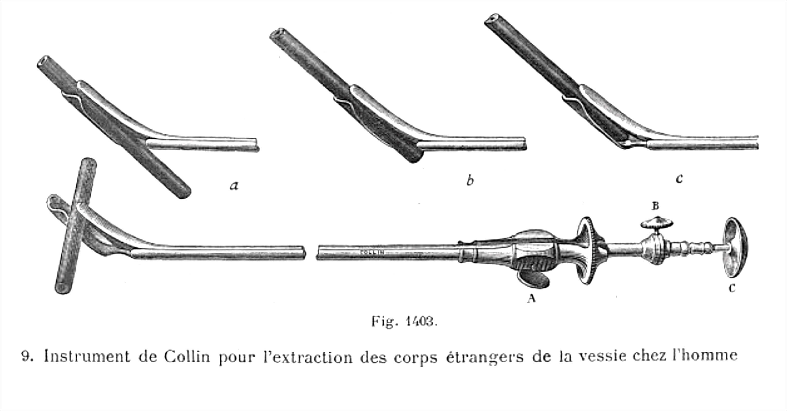
Figure 3b: Collin et Cie. Instrument Catalogue entry for the instrument, 1925.
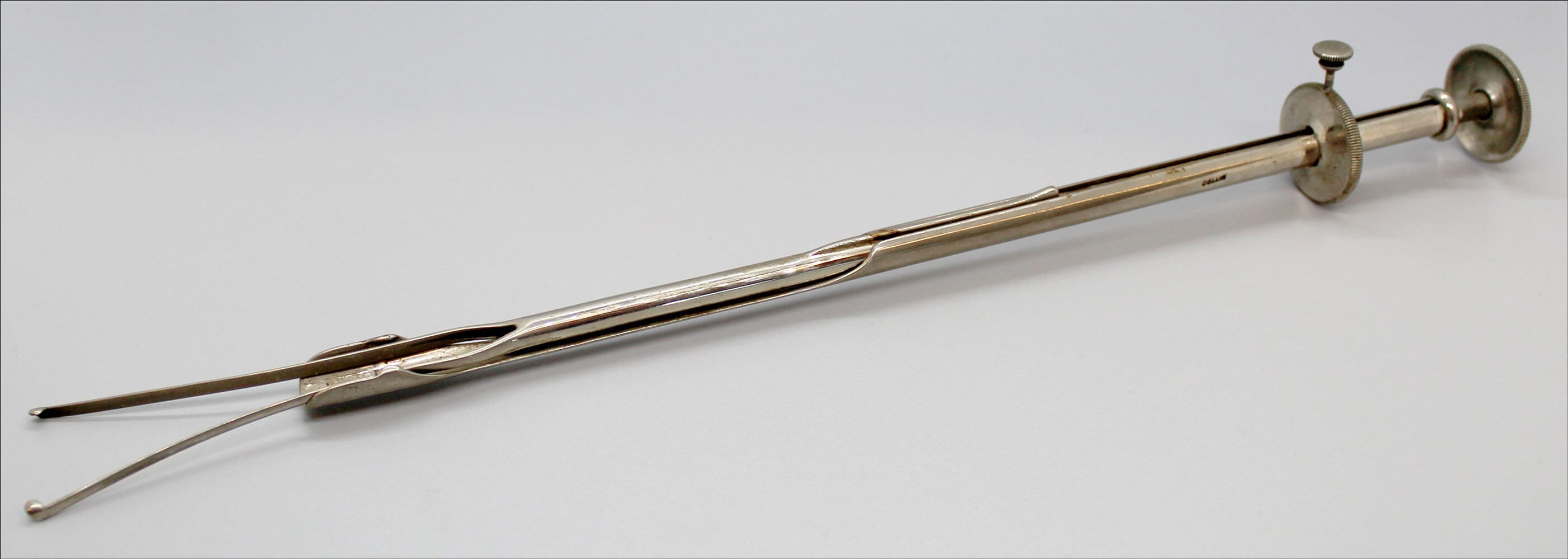
Figure 4a: Instrument for removing foreign bodies from the female bladder, Collin, Paris.
From the EAU History Office Collection.
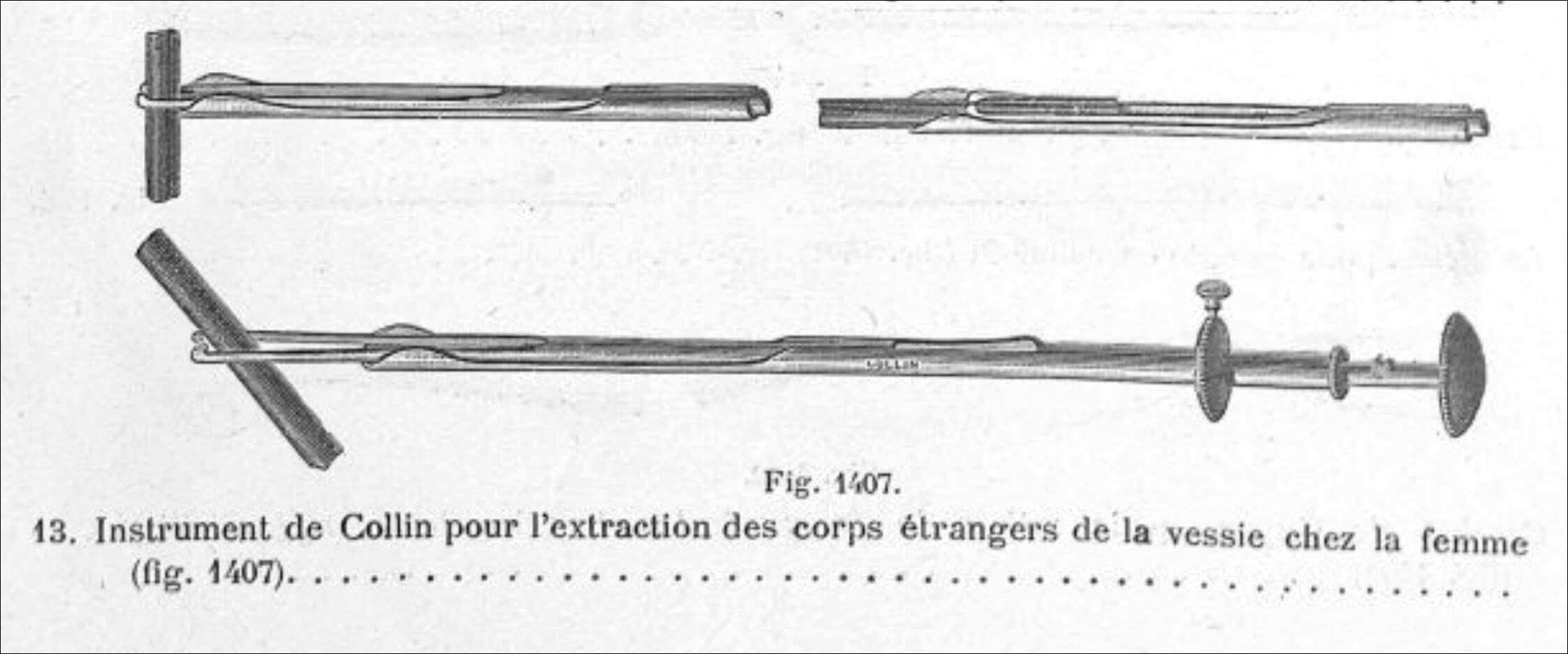
Figure 4b: Collin et Cie. Instrument Catalogue entry for the instrument, 1925.
Prior to the invention of the cystoscope, objects had to be removed blindly. Removing foreign bodies from the bladder was akin to removing small stones or stone fragments which had been created by the crushing blind lithotrites, which began to appear from the 1820s. Instruments were specifically designed to remove stone fragments; these instruments could also remove foreign bodies. Later, surgeons and instrument makers began to devise tools specifically for this. These look very similar to the instruments for removing stone fragments and indeed I imagine the two types were sometimes interchangeable (Figures 3 and 4).
The early cystoscopes were used to visualise the foreign bodies, but often, a lithotrite was then passed blindly to remove the object. Writing in 1914, Sir John Thomson Walker suggested use of a Kelly’s tube (a little like a sigmoidoscope for the urethra) in the female bladder to allow forceps or “specially constructed instruments” to grasp the objects. In the male, a combination of the Luys’ urethroscope and a lithotrite could be tried, but he felt would be less successful. Overall, he estimated that in 50% of times an open cystostomy was needed. Later, cystoscopic graspers, rongeurs, biospy forceps and optical lithotrites were used to remove the foreign bodies.
The wondrous variety of objects that have found their way into people’s bladders and the ingenious ways urologists have devised to find and remove them is a fascinating topic. Please have a look at the exhibition all about this in the Museum of Urology @ BAUS, in association with the EAU History Office:
https://www.baus.org.uk/museum/1535/instruments_for_removing_foreign_bodies