Clinical practice generates an enormous amount of patient data – histories, observations, blood results, imaging and molecular biomarkers. The problem is that these data streams rarely talk to each other.
Instead, we use them in isolation, when what we actually need is a way to merge data to guide real-time decisions and model the effects of potential treatments. Digital twins may be the tool that finally makes this possible.
What is a Digital Twin?
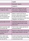
A digital twin is a patient-specific, continuously updating virtual model. It is not a risk calculator or a nomogram built to answer a single question. It integrates multiple data sources into a living virtual replica of your patient and evolves as they do [1].
It is worth distinguishing a digital twin from related concepts. Surgical simulators use standardised anatomical phantoms, while computational models lack continuous data coupling. A true digital twin is defined by three things: bidirectional data flow, real-time continuous updating and patient specificity.
The theoretical framework was proposed by Michael Grieves in a presentation at the University of Michigan in December 2002. He called it the “Mirrored Spaces Model,”, a structure comprising physical space, virtual space and bidirectional data flow between them [2]. Later, Grieves formalised this in a 2014 paper, where he adopted the term “digital twin,” a phrase coined by NASA’s John Vickers [3].
NASA demonstrated what this looked like in practice, defining a digital twin as an integrated, multiphysics, probabilistic simulation of a vehicle that uses physical models and sensor updates to mirror the life of its real-world counterpart [4]. Virtual replicas of spacecraft were continuously updated with sensor data before any real-world intervention was made. The move from aerospace to medicine became possible when high-throughput genomics, mature computational modelling and modern electronic health records converged.
The most important practical distinction is between passive and active twins. A passive twin works at a population level, useful for understanding disease dynamics, but it cannot tell you whether your specific patient needs treatment or surveillance. An active twin is different. It continuously absorbs the patient’s live data, blood results, scans, genomic markers and updates its simulations accordingly.
Beyond guidelines: the case for individual models
Clinical guidelines are an essential part of modern evidence-based medicine. They distil population-level evidence into actionable recommendations through teams of experts. But they tell us what works, on average, for a cohort of patients who met strict trial entry criteria. The patient in front of you is rarely that patient. We apply guidelines to individuals, but we rely on clinical judgement to bridge the large gap between the trials and the patient.
A digital twin inverts this logic. Rather than applying population-level conclusions downwards, it builds upwards from the individual’s own biology. It integrates their complete clinical data with the accumulated scientific evidence to generate a simulation specific to that individual.
How is a Digital Twin built?
A functional digital twin operates across five hierarchical levels, with increasing computational complexity at each stage [5].
- Level 1 (Genomic and Molecular): Integrates genetic and molecular profiles. We already engage with this in routine practice through markers like TMPRSS2-ERG fusions and BRCA mutations.
- Level 2 (Cellular): Models individual cell behaviour to predict treatment responses – for example, whether a urothelial carcinoma would respond to a PD-1 inhibitor. Cardiologists are already using this level to simulate drug-induced arrhythmias in single cells [6].
- Level 3 (Tissue and Organ): Our current clinical frontier.This level merges routine imaging – mpMRI, CT, PET – with biomechanical data to simulate organ-level tumour behaviour and surgical responses.
- Level 4 (Whole-Body): Simulates multi-system interactions simultaneously. For example, predicting how a patient’s cardiovascular, renal, and bone health will collectively respond to long-term androgen deprivation therapy.
- Level 5 (Population): Enables in silico clinical trials. Virtual control groups can accelerate drug discovery and test new therapies without exposing patients to early-phase trial risks.
Digital Twins in surgery
Surgery is one of the most compelling early environments for digital twin technology. A patient-specific virtual replica that updates dynamically during an operation, integrating live physiological data with predictive modelling, has obvious implications for surgical decision-making [7].
The Royal College of Surgeons of England projected in The Future of Surgery report that by 2035, a patient’s digital twin would continuously learn from clinical interactions, wearable devices and prescriptions to guide risk prediction and early intervention [8].
Intraoperative guidance
Intraoperative twins update in real time based on sensor data and imaging feedback. Augmented reality can overlay the virtual twin onto the surgical field, allowing surgeons to see subsurface anatomy through tissue and reduce the risk of inadvertent injury [7]. Robotic platforms are beginning to integrate these models, using the twin as a reference against which actual surgical movements are compared, enabling real-time error detection.
Postoperative monitoring
The twin’s usefulness does not end in theatre. Postoperative twins update using wearable sensors, implanted device telemetry and biomarker data to track recovery. Machine learning algorithms identify deviations from expected trajectories, thereby flagging early signs of complications before they become clinically apparent [7].
Surgical training
High-fidelity, patient-specific virtual environments allow surgeons to rehearse complex procedures on an accurate replica before any incision is made. The inclusion of bidirectional physiological coupling, trainees encounter realistic tissue responses and haptic feedback that standard VR simulators cannot replicate [7]. AI enhanced twins can analyse trainee performance and adjust scenario difficulty based on identified weaknesses.
Digital Twins in urology: where do we stand?
In practical terms, we are on the foothills of Tissue and Organ (Level 3) digital twins. The foundations are being laid out through AI augmented imaging, but the fully active urological twin is still a work in progress [9].
Prostate cancer
Diagnostic AI models are developing rapidly. The PI-CAI study – the largest of its kind, involving 10,207 MRI examinations from 9,129 patients across multiple centres – showed that a state-of-the-art AI system outperformed the mean of 62 international radiologists at biparametric MRI. It achieved a sensitivity of 0.93 against a mean radiologist sensitivity of 0.87, with only a 0.5% specificity gap versus standard clinical practice [10].
These are not digital twins, but they are the necessary precursors. The eyes of the future twin, providing the high-fidelity spatial and radiomic inputs that an organ-level model will require.
Kidney stones
While we wait for fully active twins, static patient-specific models are already proving useful in theatre. Percutaneous nephrolithotomy (PCNL) is a good example, assimilating nearly 2,000 CT images into a functional 3D mental map for access planning has always been a cognitive challenge.
A 2025 prospective randomised trial addressed this directly using immersive virtual reality (iVR) [11]. Surgeons were given an interactive 3D replica of the specific patient’s renal anatomy and surrounding structures before the procedure. They altered their planned calyx of entry in 30% of cases after exploring the model. The stone-free rate improved significantly, 33.7% versus 20.2%, and major complications (Clavien-Dindo II–IIIa) fell from 12.4% to 3.5%. The benefit was seen equally for newly qualified consultants and surgeons with 40 years of experience. These iVR models are static rather than continuously updating, they represent the Level 3 anatomical foundation on which true digital twins will be built.
Bladder cancer
Standardised reporting through VI-RADS, combined with computational imaging analysis, is building the data architecture a future digital twin will need [9]. Consider a 72-year-old with T2b/T3 bladder cancer and moderate frailty at your local cancer MDT. Currently, the choice between radical cystectomy and trimodality therapy relies on extrapolating population guidelines and a clinical assessment of frailty.
A Level 3 and 4 active twins could change this. Rather than debating subjective frailty, the team could work from a patient-specific forecast:
- Treatment response: Modelling the molecular profile predicts only a 22% probability of neoadjuvant chemotherapy downstaging, but a 65% risk of significant nephrotoxicity.
- Surgical risk: The whole-body twin integrates cardiac reserve to simulate the physiological stress of robotic cystectomy, predicting a 30% risk of Clavien-Dindo III complications and prolonged ileus.
- Radiotherapy modelling: Simulating trimodality therapy, the organ-level twin models pelvic biomechanics and predicts excellent local control with minimal radiation enteritis.
The figures above are illustrative. The architecture to generate such patient-specific predictions is the stated goal of active digital twin research but has yet to be validated clinically.
The missing link: large language models
Electronic health records are vast repositories of clinical knowledge locked inside clinic letters, histopathology reports and MDT notes. Formats that simulation engines cannot parse easily to model.
This is where large language models (LLMs) become architecturally important. They can extract structured, machine-readable information from clinical text at a scale that was previously impossible. In this configuration, the LLM acts as a bridge to structuring the patient’s history so the digital twin can run its simulations [12].
Limitations and the regulatory minefield
Digital twins require the integration of data from imaging systems, electronic health records and monitoring devices that routinely use incompatible formats and protocols [7].
Computational demands are also significant. High-fidelity physiological simulations can take hours to run, making real-time intraoperative use impractical without substantial infrastructure investment. Even static models, like the PCNL iVR replicas, require at least 90 minutes of manual segmentation to produce [11].
Validation presents a unique problem. A twin built for one individual cannot be tested against a randomised cohort in any conventional sense. When you add the risk of LLM hallucinations, which in chemotherapy protocol could be catastrophic, clinician oversight remains non-negotiable. Liability frameworks for twin-informed adverse outcomes remain legally unresolved. Regulatory frameworks including the MHRA’s AI Airlock and EU MDR 2017/745 require fundamental revision before continuously learning simulation engines can function as certified medical devices.
Conclusion
Digital twins represent the logical endpoint of precision medicine; a future where every clinical decision is grounded in that specific patient’s biology, not the average of a trial cohort or a panel consensus. The computational costs, modelling complexity, validation challenges and regulatory gaps all remain significant. The fully active whole-body twin is still a horizon rather than a destination. But the direction is clear, and the distance is closing.
References
1. Corral-Acero J, Margara F, Marciniak M, et al. The ‘Digital Twin’ to enable the vision of precision cardiology. Eur Heart J 2020;41(48):4556–64.
2. Grieves MW. PLM Initiatives [slide set]. Product Lifecycle Management Special Meeting, University of Michigan Lurie Engineering Center; 3 Dec 2002. In: Grieves M, Vickers J. Digital twin: mitigating unpredictable, undesirable emergent behavior in complex systems. In: Kahlen FJ, Flumerfelt S, Alves A (Eds.). Transdisciplinary Perspectives on Complex Systems. Cham: Springer; 2017:85–113.
3. Grieves MW. Digital twin: Manufacturing Excellence through Virtual Factory Replication (2014). White Paper.
https://www.3ds.com/fileadmin/PRODUCTS
-SERVICES/DELMIA/PDF/Whitepaper/
DELMIA-APRISO-Digital-Twin-Whitepaper.pdf
4. Glaessgen E, Stargel D. The Digital Twin Paradigm for Future NASA and U.S. Air Force Vehicles. In: 53rd AIAA/ASME/ASCE/AHS/ASC Structures, Structural Dynamics and Materials Conference. American Institute of Aeronautics and Astronautics. Honolulu, Hawaii; 2012.
5. Hernandez-Boussard T, Macklin P, Greenspan EJ, et al. Digital twins for predictive oncology will be a paradigm shift for precision cancer care. Nat Med 2021;27(12):2065–6.
6. Niederer SA, Lumens J, Trayanova NA. Computational models in cardiology. Nat Rev Cardiol 2019;16(2):100–11.
7. David-Olawade AC, Abdi AM, Amusa AO, et al. Digital twin technology in surgery: A narrative review of applications, evidence, and implementation challenges. Laparoscopic, Endoscopic and Robotic Surgery 2026. [ePub ahead of print].
8. Kerr R. The future of surgery. The Bulletin of the Royal College of Surgeons of England 2019;101(7):256–98.
9. Nedbal C, Madaan S, Nabi G, Somani BK. Digital twins in urology: a vision for the future of urological practice. Ther Adv Urol 2026;18:17562872261419592.
10. Saha A, Bosma JS, Twilt JJ, et al. Artificial intelligence and radiologists in prostate cancer detection on MRI (PI-CAI): an international, paired, non-inferiority, confirmatory study. Lancet Oncol 2024 Jul;25(7):879–87.
11. Cumpanas AD, Hernandez MC, McCormac A, et al. Preoperative Immersive Virtual Reality Applied to Percutaneous Nephrolithotomy: A Prospective Randomized Clinical Study of Surgical Planning and Clinical Outcomes. J Urol 2025;213(2):162–72.
12. Omiye JA, Gui H, Rezaei SJ, et al. Large Language Models in Medicine: The Potentials and Pitfalls: A Narrative Review. Ann Intern Med 2024;177(2):210–20.
[All links last accessed April 2026]