Case 1
- What radiological test is this and what does it show?
- What is the typical radio-nucleotide used for this study, what is its half-life and how is it excreted?
- Approximately how long does this study take to perform?
- What is the normal background radiation in the UK?
- What is the radiation exposure for this procedure?
Case 2
- What x-ray film is this and why is it different from an abdominal x-ray?
- What does the x-ray show?
- What percentage of urinary tract stones are radio-opaque and what are their likely compositions?
- What x-ray images are normally required for an IVU?
- What contrast media is required for an intravenous urogram (IVU) and what is the radiation dose of an IVU?
Case 3

- What is this study?
- What radio-isotope is used and how is it detected?
- What is the radiation dose for this study?
- How long does it take to perform this scan?
Case 4
- What imaging modality is this, what is the diagnosis?
- What is the radiation exposure for this study?
- What advantages does this study have over an IVU?
- What is the sensitivity and specificity for CT in diagnosing renal tract stones?
Radiology and imaging – answers
Case 1
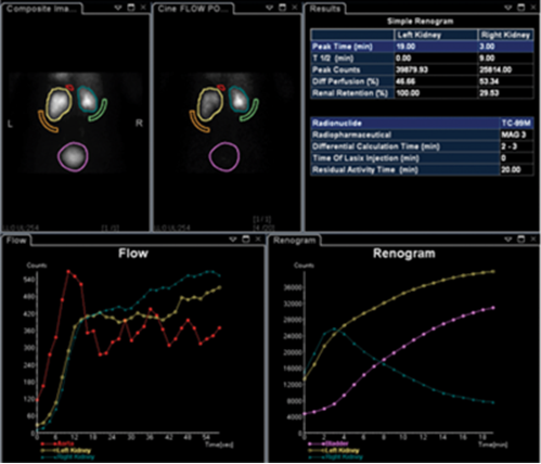
-
MAG-3 renogram, obstructed left kidney likely secondary to (pelvi-ureteric junction obstruction) PUJO.
-
MAG-3: mercaptoacetyltriglycine attached to technetium 99m, half-life six hours. 90% tubular excretion, 10% filtered at the glomerulus.
-
20-30 minutes.
-
2.5-3mSv (Higher in Aberdeen / Cornwall, approximately 8mSv).
-
Approximately 0.5-0.7mSv.
Case 2
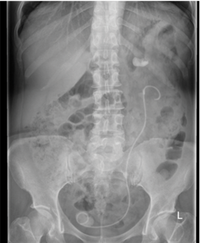
-
KUB x-ray (kidney / ureter / bladder). Film is taken at oblique angle so the patient’s pelvis is easily demonstrated / imaged, whereas an AXR is taken horizontally.
-
Left ureteric stent in-situ with calcified lower end, left mid ureteric stone and a large left upper pole stone.
-
75-85% are radio-opaque, calcium containing stones eg Calcium oxalate or calcium phosphate.
-
Plain KUB, immediate nephrogram, five minute film, 10 minutes, 20 minutes and a post-micturition study +/- delayed films if required. 5. Non-ionic, low-osmolality contrast agent (e.g. Omnipaque 1ml/Kg), 2.5mSv.
Case 3
-
Radionuclide bone scan.
-
Technetium 99m, Gamma camera.
-
6.5mSv.
-
2-4 hours
Case 4
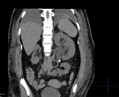
-
Plain CT KUB, coronal reconstruction demonstrating left PUJ stone with associated hydronephrosis.
-
4.5mSv
-
Quick scan (20-30 seconds), easy to interpret, no IV contrast needed, can establish differential diagnosis.
-
Sensitivity >95%, specificity >96%.


