Case 1
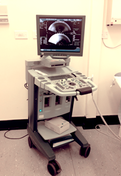
- Name the machine shown in the picture and describe how it works.
- What is the standard frequency of ultrasound waves in this machine?
- What frequency of ultrasound waves are utilised in an abdominal and scrotal ultrasound?
Case 2
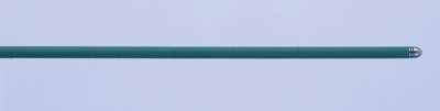
Figure 1.
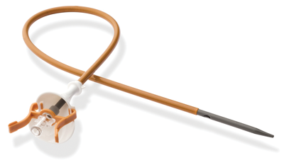
Figure 2.
- What is shown in Figures 1 and 2?
- Describe the standard diameter and length of Figure 1.
- What material are such wires constructed from?
- Describe the different variations in these wires.
- What size and length can Figure 2 be?
Case 3
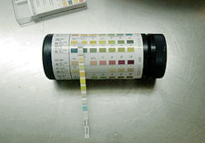
- What is shown in the figure and what does it measure?
- How does the test for haemoglobin work?
- Name potential false positives and false negatives for haemoglobin testing.
- How does the nitrite test work?
Technology – answers
Case 1
-
Transrectal ultrasound machine. It works by using high frequency sound waves which are produced by the passage of current through a piezoelectric transducer.
-
Transrectal ultrasound probe works at about 7.5MHz.
-
Abdominal ultrasound is 3-5MHz, scrotal ultrasound is 10MHz. Lower frequencies have deeper penetration; higher frequencies result in more superficial penetration but increased resolution.
Case 2
-
Figure1: standard ureteric guide wire. Figure 2: ureteric access sheath.
-
0.035-0.038 inches in diameter and approximately 150cm long.
-
Stainless steel core with a polytetrafluoroethylene (PTFE) coating.
-
They can come as a standard PTFE wire throughout, a hydrophilic tip and Nitinol/PTFE shaft, completely hydrophilic throughout or as a stiff wire. Other options include a curved hydrophilic tip and dual tipped wires (hydrophilic one end and standard the other).
-
Ureteric access sheaths come in a variety of sizes (9/11Fr to 13/15Fr) and lengths (28cm-55cm).
Case 3
-
The picture shows a multistix urine test strip. It measures: pH, haemoglobin, nitrites, leucocytes, protein, glucose, ketones, specific gravity, urobilinogen.
-
It is based on the alteration of a chromogen (orthotolidine) by the peroxidase activity of haemoglobin, which results in a colour change (to a blue colour).
-
False positives
• Myoglobinuria
• Oxidising agents
• Peroxidases False negatives
• Ascorbic acid (Vitamin C)
• High gravity content of the urine
-
Gram negative bacteria, which are commonly found in the urinary tract, release urease which convert nitrates (normally present in the urine) to nitrites which are not normally found in the urine. Nitrites then react with the aromatic amine reagent on the dipstick to form a diazonium salt. This salt then interacts with hydroxybenzoquinolone to form a pink colour.
Further reading
Arya M, Shergill IS, et al. Viva Practice for the FRCS (UROL) Examination. Oxford, UK; Radcliffe Publishing LTD; 2010.
For Part 1 of this topic click here.
For Part 3 of this topic click here.