Spot Tests
Advanced renal tumours
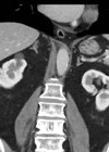
Case 1 A 67-year-old gentleman underwent a CT scan after presenting with visible haematuria and weight loss. His comorbidities include hypertension, type II diabetes mellitis and hypercholesterolaemia. He is a smoker. Figure 1. Figure 2. What do Figures 1 and...
Localised renal cancer
Case 1 A 56-year-old lady is referred to the urology clinic after the GP conducted an ultrasound abdomen for deranged liver function tests and found a renal lesion. She is otherwise fit and well. Figure 1. What is the sensitivity...
Prostate cancer management 2 – metastatic disease
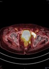
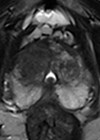
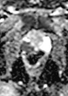
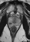
A 72-year-old gentleman is referred to you in the two-week wait clinic with a prostate specific antigen (PSA) of 22ug/L. He is otherwise fit and well and does not take any regular medication. His multi-parametric magnetic resonance imaging (mpMRI) shows...
Prostate cancer management 1 – non-metastatic disease
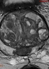
You are referred a 68-year-old gentleman to the rapid access prostate clinic with a serum prostate specific antigen (PSA) of 12ug/L. He is otherwise fit and well with mild voiding lower urinary tract symptoms (LUTS). He undergoes a multi parametric...
Prostate cancer series: diagnostics 2
- Click here for Part 1 - A 68-year-old male was referred to the two-week wait prostatic clinic with a serum prostate specific antigen (PSA) of 17. He had no bothersome lower urinary tract symptoms, relevant past medical history or...
Prostate cancer series: diagnostics 1
- Click here for Part 2 - A 58-year-old male is referred to your rapid access prostate clinic with a prostate specific antigen (PSA) of 6.0ng/ml. He has no lower urinary tract symptoms (LUTS), past medical history (PMH), or family...
The role of embolisation in urology
Case 1 An 86–year–old male presented with visible haematuria and suprapubic pain. He had a history of diabetes, heart failure, benign prostatic hypertrophy, aortic valve replacement, deep vein thrombosis (DVT) and atrial fibrillation (AF) and was anticoagulated on a non-VKA...
Non-urothelial bladder malignancies
Case 1 An 80-year-old gentleman presented with a history of visible haematuria and recurrent urinary tract infections (UTIs). He has been performing intermittent self catheterisation (ISC) for detrusor underactivity for over 20 years. A flexible cystoscopy showed these appearances of...
Renal masses
Case 1 A 70-year-old female presented under the medical team with malaise, weight loss, and deranged liver function tests (LFTs) and calcium (ALP 350, GGT 650, Serum bilirubin 29, normal aminotransferases, Ca 3.3). An abdominal ultrasound scan (USS) was performed...
Prostate cancer
Case 1 A 65-year-old man is referred to your two-week wait (2WW) clinic with a PSA of 7.0ng/mL. He has no lower urinary tract symptoms (LUTS), no past medical history, no family history of prostate cancer (PCa) and his performance...
Penile cancer
Case 1 A 67-year-old man presents with a worsening red patch over the past three months. It looks velvety in some areas. What is the most likely diagnosis? What are the risk factors? How do you classify this condition? How...
Upper urinary tract urothelial cell carcinoma
Case 1 A 64-year-old man presents to the haematuria clinic with visible haematuria, on a background of a 40 pack-year smoking history and family history of bowel cancer in his sister at the age of 48. A CT was performed...


