Bladder cancer (BCa), ranking as the 10th most common cancer worldwide, poses a significant health burden with high morbidity and mortality [1]. Timely tumour detection and accurate evaluation are crucial for effective management, as the prognosis is dependent on the stage and grade at diagnosis [2].
While conventional diagnostic methods such as cystoscopy, imaging, and histopathology remain the keystone of bladder cancer diagnostics, they are often subject to interobserver variability and limitations in accuracy [3]. Emerging breakthroughs in artificial intelligence have granted opportunities to overcome these limitations to facilitate diagnostic precision.
Terms and definitions:
Artificial intelligence (AI) refers to advanced computer systems designed to mimic human cognitive processes, including reasoning, decision-making, and executive functions, by utilising complex, non-linear mathematical algorithms to achieve optimal outcomes [4]. Urology, traditionally at the forefront of technological advancements, particularly in robotic surgery, has now embraced AI to enhance radiographic analysis, histopathological evaluation, diagnostic accuracy, and the prediction of prognostic outcomes based on diverse, patient-specific variables [5]. This article aims to explore the emerging role of AI in BCa diagnostics. To provide clarity, key terms essential for understanding this rapidly evolving field are defined in this section.
Machine learning (ML): This is a subdivision of AI that employs data to learn task performance through various statistical algorithms, instead of being programmed with pre-determined instructions and conditions. ML analyses diverse data formats such as images, numerical data, examples, and prior experiences through processes that include training, validation, and testing across various datasets [4,6].
Most ML operations fall under supervised learning, where human specialists provide labelled datasets, often including annotations like ‘benign’ or ‘malignant’ in the context of bladder lesions. Consequently, seamless collaboration between ML experts and medical researchers is crucial to applying ML effectively in clinical settings [7].
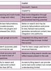
Deep learning (DL), a specialised branch of ML, employs deep artificial neural networks (ANNs) designed to mimic the structure and function of the human nervous system [8]. ANNs consist of multiple interconnected layers that enable the automated learning and processing of complex, large-scale datasets [9]. Within ANNs, convolutional neural networks (CNNs) are particularly noteworthy for their ability to replicate and reuse learned neuronal patterns across various regions, allowing for efficient processing and recognition of visual and spatial patterns. This makes CNNs highly effective in applications such as image and pattern recognition, commonly used in radiological, endoscopic, and histopathological analyses for medical diagnostics, including BCa detection [10]. A simplified representation of this framework is illustrated in Figure 1.

Figure 1: Simplified framework of artificial intelligence employed in medicine.
AI in bladder cancer diagnostics
BCa diagnostics involves a series of investigative steps aimed at detecting tumours, followed by accurate grading and staging to guide treatment (Figure 2). AI has been progressively integrated into each phase with an aim to enhance diagnostic precision and accelerating processes across various diagnostic modalities.

Figure 2: Overview of bladder cancer diagnostics.
Bladder tumour detection
White light cystoscopy remains the standard method for detecting bladder tumours despite its significant diagnostic error rates, which range from 10% to 40% [11,12]. These error rates were addressed with additional features like blue light cystoscopy and narrow band imaging. However human errors caused by visual and mental fatigue can still exist. Given that AI can mitigate human error, integrating AI tools alongside clinician expertise has proven effective in enhancing diagnostic accuracy. One such tool is CystoNet, developed by Shkolyar et al., which utilises a CNN algorithm for tumour detection. Trained on a dataset comprising images of normal bladder epithelium and papillary urothelial carcinoma, CystoNet distinguishes between malignant and benign images with a sensitivity of 91% and a specificity of 99% [13].
The Cystoscopy Artificial Intelligence Diagnostic System (CAIDS) is another prominent example, showcasing exceptional diagnostic accuracy and sensitivity. In a multicentre study, CAIDS achieved a diagnostic accuracy of 0.977 in internal validation and 0.990 in external validation, surpassing expert urologists in both precision and speed. Notably, the system excels in detecting complex lesions, including flat cancerous tissues such as carcinoma in situ, which are typically challenging to identify [14].
DL and CNN models have also significantly improved BCa detection using urinary cytology and imaging methods. Notable studies include those by Nojima et al., who employed a 16-layer visual geometry group CNN with high accuracy in identifying malignant lesions (area under the curve (AUC) 0.9890), while Awan et al. found Xception effective in identifying atypical cells (AUC 0.99). Vaickus et al. achieved 95% accuracy in automating the Paris System for cell typing. Sanghvi et al. validated a CNN model achieving an AUC of 0.88 for high-grade BCa. Methods like atomic force microscopy and focal loss-enhanced DL models further demonstrated promising diagnostic accuracy, reaching up to 94%.
Urine metabolomics also offers promise in bladder cancer detection. Shao et al. identified imidazoleacetic acid as a marker using an ML decision tree with 76.6% accuracy. Kouznetsova et al. highlighted biomarkers like D-glucose and glycerol, achieving 82.54% accuracy, emphasising metabolomics’ potential for early and late-stage BCa detection [15].
Bladder segmentation research in BCa imaging
Bladder segmentation which aims to differentiate bladder from surrounding structures on imaging is a critical initial step in developing computer-aided diagnosis for BCa. However, it is challenging due to the bladder’s variable anatomy in size and shape, and the low-contrast boundaries between the bladder wall and surrounding soft tissue. Inaccurate segmentation can lead to false negatives by excluding tumours within the bladder or false positives by including non-bladder structures.
Cha et al. developed a CNN algorithm to recognise bladder patterns in CT urography scans. This model produced a bladder likelihood map fed into a level-set segmentation method, achieving a Jaccard index of 0.76, measuring overlap accuracy.
Subsequently, Ma et al. trained an end-to-end U-net model on the same dataset, eliminating the need for user-provided regions of interest (ROIs) or auxiliary level-set post-processing. This is a DL model with a U-shaped architecture, that improved the Jaccard index to 0.85, showing enhanced segmentation performance.
Similar DL models have also been successfully applied to bladder segmentation in MRI scans, advancing the accuracy and reliability of BCa imaging [16].
Tumour grading
Grading assesses the degree of tumour cell abnormality, distinguishing between low-grade and high-grade cancers, a critical distinction for evaluating a tumour’s potential to invade surrounding tissues, metastasize, and recur. Efforts to fully automate tumour grading using AI have incorporated advanced techniques such as MRI radiomics [16]. Jansen et al. employed a U-net segmentation network, achieving grading accuracy of 76% for low-grade and 71% for high-grade bladder urothelium cancers. Similarly, Wang et al. and Zhang et al. developed deep learning (DL) systems using MRI data, achieving an area under the curve (AUC) exceeding 86% [15]. However, these studies were limited by small sample sizes, reducing their immediate clinical applicability.
Recent advancements have focused on training AI models with larger datasets to improve reliability. Pan et al. introduced a pathological artificial intelligence diagnostic model (PAIDM) to identify and grade tumour histopathology specimens. This model demonstrated an AUC of 85%, outperforming junior pathologists, compared to intermediate pathologists while being slightly less accurate than senior pathologists, who achieved an average of 92.5% but required more time for diagnosis. The PAIDM is anticipated to serve as a valuable adjunct in both clinical and training settings [17].
Shalata et al. have advanced non-muscle invasive bladder cancer (NMIBC) grading by using a ShuffleNet-based AI system, a multi-scale CNN. This model addressed biases and subjectivity inherent in pathology, achieving an accuracy of 94.25%, sensitivity of 94.47%, and specificity of 94.03%. Future efforts aim to enhance the generalisability of these results through multi-institutional datasets and include a broader range of pathological subtypes [18].
Tumour staging
Staging BCa into muscle-invasive bladder cancer (MIBC) and NMIBC categories is a critical step in guiding treatment decisions. A growing body of literature highlights the accuracy and effectiveness of AI in staging BCa. Imaging-based AI models, such as CNNs with support vector machines (SVMs), leverage features from multiparametric MRIs using the VI-RADs (The Vesical Imaging- Reporting and Data System) and CT scans for tumour classification [15]. Li et al. enhanced feature selection by incorporating advanced algorithms like recursive feature elimination and least absolute shrinkage and selection operator (LASSO). Their multi-task model demonstrated superior performance over radiomics and single-task models, achieving an AUC of 93.2% and significantly streamlining the process of determining muscle invasion using T2-weighted images [19].
Histopathological analysis is a key component of BCa staging. Yin et al. utilised AI systems to analyse haematoxylin and eosin (H&E) slides, employing classifiers such as SVMs and random forest to differentiate between Ta and T1 BCa stages. These models, trained with pathologist-labelled data, achieved classification accuracies ranging from 91% to 96% by automating the detection of subtle patterns indicative of tumour invasiveness. In contrast, unsupervised clustering analysis, where the AI system was not explicitly trained, struggled to distinguish between Ta and T1 stages. This limitation likely arises from the microscopic subtlety of the differences between these stages, which even advanced algorithms cannot reliably discern without supervised training [20].
Conclusion
AI holds significant promise as a complementary tool in BCa diagnostics, grading, and staging. However, its integration into clinical practice requires prospective real-time validation through larger, multi-institutional datasets to ensure generalisability and reliability. While AI enhances precision and efficiency, it must work alongside human expertise, emphasising collaboration for accurate, tailored and holistic patient care.
TAKE HOME MESSAGES
-
AI enhances bladder cancer detection: AI, particularly DL and CNN models, has demonstrated high accuracy in BCa detection through tools like CystoNet, significantly improving diagnostic precision and mitigating human error in cystoscopy.
-
AI improves tumour grading: AI models, such as U-net and ShuffleNet-based systems, have shown promise in automating tumour grading by distinguishing between low-grade and high-grade cancers, enhancing diagnostic accuracy, and addressing subjectivity inherent in traditional pathology methods.
-
AI supports tumour staging: Advanced AI systems utilising imaging and histopathological data, including CNNs, SVMs, and LASSO algorithms, enable accurate staging, aiding critical treatment decision-making.
-
Integration of AI in imaging: AI applied to bladder segmentation in CT and MRI scans improves the accuracy and efficiency of tumour detection by overcoming challenges related to low-contrast boundaries and anatomical variability.
-
To enable real-time use of the existing and new AI tools in BCa diagnostics, multicentric studies with larger datasets are essential. To ensure accountability and diagnostic accuracy, AI can only serve as an adjunct to clinician’s expertise which is non-negotiable.
References
1. Sung H, Ferlay J, Siegel RL, et al. Global Cancer Statistics 2020: GLOBOCAN Estimates of Incidence and Mortality Worldwide for 36 Cancers in 185 Countries. CA Cancer J Clin 2021;71(3):209–49.
2. Babjuk M, Burger M, Capoun O, et al. European Association of Urology Guidelines on Non-muscle-invasive Bladder Cancer (Ta, T1, and Carcinoma in Situ). Eur Urol 2022;81(1):75–94.
3. Rossin G, Zorzi F, Ongaro L, et al. Artificial intelligence in bladder cancer diagnosis: current applications and future perspectives. BioMedInformatics 2023;3(1):104–14.
4. Russell S, Norvig P. Artificial Intelligence: A Modern Approach. 2016; Pearson.
5. Chen AB, Haque T, Roberts S, et al. Artificial intelligence applications in urology: reporting standards to achieve fluency for urologists. Urol Clin North Am 2022;49(1):65–117.
6. Alpaydin E. Introduction to Machine Learning. 2020; MIT Press.
7. Rajkomar A, Dean J, Kohane I. Machine learning in medicine. N Engl J Med 2019;380(14):1347–58.
8. LeCun Y, Bengio Y, Hinton G. Deep learning. Nature. 2015;521(7553):436–44.
9. Goodfellow I, Bengio Y, Courville A. Deep Learning. 2016; MIT Press.
10. Litjens G, Kooi T, Bejnordi BE, et al. A survey on deep learning in medical image analysis. Medical Image Analysis 2017;42:60–88.
11. Kata SG, Zreik A, Ahmad S, et al. Concurrent bladder cancer in patients undergoing photodynamic diagnostic ureterorenoscopy: how many lesions do we miss under white light cystoscopy? Cent European J Urol 2016;69(4):334–40.
12. Ren H, Park KC, Pan R, et al. Early detection of carcinoma in situ of the bladder: a comparative study of white light cystoscopy, narrow band imaging, 5-ALA fluorescence cystoscopy and 3-dimensional optical coherence tomography. J Urol 2012;187(3):1063–70.
13. Shkolyar E, Jia X, Chang TC, et al. Augmented bladder tumor detection using deep learning. European Urology 2019;76(6);714–18.
14. Wu S, Chen X, Pan J, et al. An artificial intelligence system for the detection of bladder cancer via cystoscopy: a multicenter diagnostic study. J Natl Cancer Inst 2022;114(2):220–7.
15. Ferro M, Falagario UG, Barone B, et al. Artificial intelligence in the advanced diagnosis of bladder cancer-comprehensive literature review and future advancement. Diagnostics 2023;13(13):2308.
16. Borhani S, Borhani R, Kajdacsy-Balla A. Artificial intelligence: a promising frontier in bladder cancer diagnosis and outcome prediction. Crit Rev Oncol Hematol 2022;171:103601.
17. Pan J, Hong G, Zeng H, et al. An artificial intelligence model for the pathological diagnosis of invasion depth and histologic grade in bladder cancer. Jour Transl Med 2023;21(1):42.
18. Shalata AT, Alksas A, Shehata M, et al. Precise grading of non-muscle invasive bladder cancer with multi-scale pyramidal CNN. Scientific Reports 2024;14(1):25131.
19. Li J, Qiu Z, Cao K, et al. Predicting muscle invasion in bladder cancer based on MRI: A comparison of radiomics, and single-task and multi-task deep learning. Comput Methods Programs Biomed 2023;233:107466.
20. Yin P, KC K, Wei S, et al. Histopathological distinction of non-invasive and invasive bladder cancers using machine learning approaches. BMC Med Inform Decis Mak 2020;20:162.
Declaration of competing interests: None declared.