The Meyer-Weigert rule is a fundamental anatomical principle in urology that describes the typical orientation of ureteral orifices in duplex kidneys. In a duplex system, the upper pole ureter usually inserts inferomedially, while the lower pole ureter inserts superolaterally.
This anatomical variation can complicate diagnostic and therapeutic interventions, particularly in emergency settings such as ureteric stenting for obstructing ureteric stones. Missing a duplex ureter during such procedures can lead to incomplete treatment, persistent symptoms, and potential complications. This literature review explores the challenges of identifying duplex ureters, the implications of missed diagnoses, and strategies to improve detection and management.
Anatomy and embryology of duplex ureters
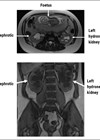
A duplex ureter, or duplex collecting system, is a congenital anomaly resulting from incomplete fusion of the ureteric buds during embryogenesis. It is one of the most common renal anomalies, occurring in approximately 0.8% of the population [1]. The Meyer-Weigert rule provides a predictable pattern for the insertion of duplex ureters into the bladder: the upper pole ureter drains the lower renal moiety and inserts inferomedially, while the lower pole ureter drains the upper renal moiety and inserts superolaterally [2]. This anatomical arrangement is critical for understanding the clinical presentation and management of duplex systems (Figure 1).

Figure 1: An anatomical drawing of complete unilateral duplication of the right ureter with ectopic orifice into the prostatic urethra. LU - lower ureter, UU - upper ureter, B – urinary bladder. CC BY 4.0 reproduced from: Balawender K, Wawrzyniak A, Pliszka A, et al [11].
Challenges in diagnosing duplex ureters
Diagnosing a duplex ureter can be challenging, especially in emergency settings where time and resources are limited. Imaging modalities such as ultrasound, intravenous urography (IVU), and computed tomography (CT) are commonly used to evaluate ureteric stones. However, these techniques may not always clearly delineate a duplex system, particularly if the anomaly is not suspected or if the imaging is focused solely on the obstructing stone [3]. In some cases, the duplex ureter may be overlooked due to overlapping structures or inadequate contrast opacification.
Case reports and clinical implications
Several case reports highlight the challenges and consequences of missing a duplex ureter during emergency ureteric stenting. For example, a study by Gunawardena [4] described a patient with a duplex ureter who presented with flank pain and was initially diagnosed with a single obstructing ureteric stone. During ureteric stenting, only one ureter was cannulated, leaving the obstructed duplex ureter untreated. The patient subsequently developed persistent symptoms and required a second procedure to address the missed ureter. This case underscores the importance of considering anatomical variations during diagnostic and therapeutic interventions.
Similarly, a report by Ntalianis [5] emphasised the role of preoperative imaging in identifying duplex systems. In their case, a patient with a known duplex ureter underwent ureteric stenting for an obstructing stone. However, the stent was inadvertently placed in the non-obstructed ureter, leading to incomplete relief of symptoms. The authors highlighted the need for careful review of imaging studies and intraoperative confirmation of ureteral anatomy to avoid such errors.
Diagnostic strategies
To improve the detection of duplex ureters, several diagnostic strategies have been proposed. CT urography (CTU) is considered the gold standard for evaluating ureteral anatomy, as it provides high-resolution images of the urinary tract and can clearly delineate duplex systems [6]. In cases where a duplex ureter is suspected, CTU should be performed to confirm the diagnosis and guide treatment planning.
Additionally, cystoscopy and retrograde pyelography (RGP) are valuable tools for identifying duplex ureters during ureteric stenting. Cystoscopy allows direct visualisation of the ureteral orifices, while RGP provides detailed images of the ureteral anatomy. These techniques can help confirm the presence of a duplex system and ensure accurate stent placement [7].
Management considerations
When a duplex ureter is identified, management strategies should be tailored to the specific clinical scenario. In cases of obstructing ureteric stones, both ureters may require stenting to ensure complete drainage and relief of symptoms. If only one ureter is stented, the patient may experience persistent obstruction and complications such as hydronephrosis, infection, or renal impairment [8].
In some cases, surgical intervention may be necessary to address underlying anatomical abnormalities or recurrent stone disease. Ureteroureterostomy or ureteral reimplantation may be considered to correct ureteral duplication and prevent future complications [9,10]. However, these procedures should be performed by experienced urologists to minimise the risk of complications.
Conclusion
The Meyer-Weigert duplex ureter is a clinically significant anatomical variation that can complicate the management of ureteric stones, particularly in emergency settings. Missing a duplex ureter during ureteric stenting can lead to incomplete treatment, persistent symptoms, and potential complications. To improve outcomes, clinicians should maintain a high index of suspicion for duplex systems, utilise advanced imaging techniques such as CTU, and employ intraoperative confirmation of ureteral anatomy. By adopting these strategies, the risk of missed diagnoses can be minimised, and patients can receive optimal care.
References
1. Whitten SM, Wilcox DT. Duplex systems. Prenat Diagn 2001;21(11):952–7.
2. Darr C, Krafft U, Panic A, et al. Renal duplication with ureter duplex not following Meyer-Weigert-Rule with development of a megaureter of the lower ureteral segment due to distal stenosis – a case report. Urol Case Rep 2019;28:101038.
3. Nyanhongo DK, Antil S, Nasir S. Pitfalls of the duplex system: the mystery of the missing stone. BMJ Case Rep 2017;2017:bcr2017220198.
4. Gunawardena S, Ranasinghe WKB, McCahy P. Beware the stone in the duplex: Use of CT intravenous pyelogram (CT IVP) in detecting calculi in duplex ureteric systems. J Clin Urol 2016;9:128–30.
5. Malligiannis Ntalianis K, Cheung C, Resta C, et al. Unilateral duplicated collecting system identified during pelvic lymphadenectomy: a case report and literature review. Cureus 2022;14(6):e26331.
6. Dervishi B, Hyseni F, Musa J, et al. The importance of CT urography in early diagnosis of anatomical variations in urogenital tract: case presentation. Radiol Case Rep 2022;17(10):4025–9.
7. Share JC, Lebowitz RL. The unsuspected double collecting system on imaging studies and at cystoscopy. AJR Am J Roentgenol 1990;155(3):561-4.
8. Adenipekun A, Gaballa N, Darrad M. Ureteric calculus in a left complete duplex system masquerading as an impacted stone: a case report and literature review. Cureus 2023;15(7):e41489.
9. Wu C, Ji F, Zhang H, et al. Treatment for complete bilateral duplex kidneys with severe hydronephrosis and ureterectasis of the upper moiety in a child: a case report and literature review. Front Surg 2022;9:1019161.
10. Chacko JK, Koyle MA, Mingin GC, Furness PD. Ipsilateral ureteroureterostomy in the surgical management of the severely dilated ureter in ureteral duplication. Journal of Urology [Internet] 2007;178(4S):1689–92.
11. Balawender K, Wawrzyniak A, Pliszka A, et al. Ectopic ureter: A concise narrative review with anatomical and clinical commentaries. Translational Research in Anatomy 2022;29:100220.
Declaration of competing interests: None declared.