Clear cell urothelial carcinoma (CCUC) is a rare morphological variant of transitional cell carcinoma (TCC). It can occur anywhere along the urothelial tract and is characterised histologically by high grade carcinoma with an abundance of clear, glycogen-rich cytoplasm [1].
Alternative diagnoses for such tumours occurring within the bladder include clear cell adenocarcinoma, metastatic renal cell carcinoma (RCC) or advanced prostatic adenocarcinoma. Clear cell adenocarcinoma of the bladder typically occurs in women, with histology demonstrating a combination of solid, papillary and tubulocystic architecture; papillae lined by a single layer of epithelium; eosinophilic or clear cytoplasm and a hobnailed appearance to cells. There may be positivity for cytokeratin 7 and CA-125, with variable positivity for cytokeratin 20 on immunohistochemistry. Metastatic RCC is usually excluded by the presence of a renal mass and multiorgan metastases. Histologically, there is an alveolar or nested pattern with intervening septa containing capillaries and lymphocytes. Immunohistochemistry demonstrates cytokeratin, vimentin, RCC antigen, PAX-2 and no reaction for cytokeratin 7 and 20. Prostatic adenocarcinoma tends to involve the muscularis propria, sparing lamina propria and urothelium as there is direct invasion of bladder wall from the prostate. Despite high grade and stage disease, nuclei tend to appear more homogenous with pleomorphism and high mitotic rate being uncommon. Strong reaction with PSA or PSAP supports the diagnosis immunohistochemically.
Evidence of this rare variant’s incidence and natural history is limited to case reports or small series [2-21]. A total of 31 cases of CCUC arising in the bladder has been reported in the published literature [2-16]. Patients have typically presented with visible haematuria and a background history of smoking. Other modes of presentation included progressive lower urinary tract symptoms, urinary retention and incidental identification of the tumour on imaging. Up front treatment modalities have primarily involved radical cystectomy or transurethral resection of bladder tumour (TURBT). Where histological findings have been reported, high grade disease was demonstrated in all cases. Muscle-invasive tumours were present in 26/31 cases (84%) and nodal or distant metastasis were identified in 10/31 (32%). Follow-up ranged from three months to four years, in which time recurrence occurred in 3/31 (10%) and mortality in 13/31 (42%). Claps et al. reported outcomes from a multicentre retrospective cohort totalling 1082 patients [16]. All cases had undergone upfront radical cystectomy and bilateral pelvic lymph node dissection for T1-T4 N0 M0 TCC of the bladder. They compared disease specific survival in patients with variant histology and showed that CCUC demonstrated the worst survival (HR 10.8, 95% CI 1.42–81.4 [p=0.021]) among patients with organ-confined disease.
Upper tract CCUC appears to be a rarer entity, with only eight cases previously reported in the literature [3,12,17-21]. Among these, presenting features included flank pain and visible haematuria with a palpable mass identified on examination in one case. Computed tomography (CT) showed evidence of an upper tract mass with anatomical sites including the renal pelvis and upper ureter. Radical nephroureterectomy was performed in six patients (75%), nephrectomy and upper ureterectomy in one (13%) and radical nephrectomy in one (13%). Stage of disease was reported in four patients as Ta-T3 with lymph node and distant metastasis present in one case (13%). Follow-up time was 1-19 months with one recurrence occurring at six months (13%) and two deaths (25%) within six months.
We report our experience of two cases which demonstrate the aggressive natural history of CCUC. The first patient presented with disease arising from the bladder while the second case featured an upper tract tumour.
![]()
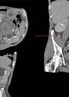
Figure 1: Axial CT section demonstrating upper tract CCUC recurrence in left retroperitoneum.
![]()
Figure 2: Coronal CT section of upper tract CCUC recurrence.
Case 1
A 64-year-old female patient with a background of previous stroke, hypertension and raised body mass index (BMI) underwent staging CT for a newly diagnosed anal cancer. This incidentally identified an anterior bladder wall lesion, for which TURBT was performed at her local hospital. Histology demonstrated at least pT1 CCUC with carcinoma-in-situ (CIS), though the specimen lacked muscularis propria. Re-resection was performed three months later but this was non-diagnostic. She completed a course of chemoradiotherapy for anal cancer, which was staged as T4 N1a squamous cell carcinoma.
The patient was referred to our department one year following initial diagnosis due to her anal cancer being treated at our centre by colorectal surgery. She had declined surveillance cystoscopies in that time. Check flexible cystoscopy was performed which demonstrated no visible pathology within the bladder or urethra. Repeat cystoscopy three months later identified widespread cystitis cystica but no concerning abnormalities on white light or narrow band imaging. Surveillance CT chest / abdomen / pelvis at that time, however, demonstrated multiple liver metastases which ultrasound-guided biopsy identified as high-grade urothelial carcinoma.
Palliative chemotherapy was commenced but her course stopped prematurely due to fatigue and diarrhoea. Two further check cystoscopies have demonstrated stable, unremarkable appearances of the urothelium. The patient continues to be followed up with regular surveillance cystoscopies by urology and is awaiting discussion with oncology regarding reduced dose chemotherapy.
Case 2
A 72-year-old female patient with a previous history of myocardial infarction, type two diabetes, hypertension and rheumatoid arthritis underwent CT chest / abdomen / pelvis to investigate weight loss and lethargy. This demonstrated a 34mm left-sided renal lesion with a distended renal pelvis and three enlarged para-aortic nodes, the largest of which was 21mm. Radiological staging was T3 N1 M0. Flexible cystoscopy identified no lesion within the urethra or bladder and positron emission tomography (PET) CT confirmed the presence of left-sided retroperitoneal lymphadenopathy with additional positive nodes demonstrated.
Given the extent of retroperitoneal lymph node involvement, the tumour was considered unresectable and the patient was offered ureteroscopic biopsy with a view to undergoing systemic oncological therapy. She instead sought private treatment at another centre where robotic left nephroureterectomy and lymph node dissection was performed, revealing grade 3 pT3 pN0 R0 clear cell TCC of the left renal pelvis with lymphovascular invasion. Pre- and para-aortic lymph nodes were negative for malignancy.
She re-attended via the emergency department two months postoperatively with left flank pain and significantly raised inflammatory markers. A left-sided 8.3 x 6.5cm retroperitoneal mass with new para-aortic lymphadenopathy was identified on CT abdomen / pelvis and was diagnosed as recurrent disease (Figures 1 and 2). This was engulfing the descending colon and extended inferiorly to psoas. She was treated with intravenous antibiotics and reviewed by oncology but deemed too unfit for systemic oncological treatment in the context of her acute illness. She died three months after her operation.
TAKE HOME MESSAGE
-
Clear cell urothelial carcinoma is a rare and aggressive morphological variant of TCC which can occur throughout the urinary tract.
-
Tumour grade and stage tend to be high at presentation.
-
There is high risk of early recurrence and mortality despite aggressive treatment.
-
Clinical management should focus on rapid diagnosis and radical treatment at an early stage.
-
Radical treatment should take the form of radical cystectomy for CCUC of the bladder and radical nephroureterectomy in upper tract CCUC.
-
Consideration should be given to adjuvant treatment modalities given the high incidence of recurrence and mortality despite radical therapy.
References
1. Amin MB. Histological variants of urothelial carcinoma : diagnostic , therapeutic and prognostic implications. Modern Pathology 2009;22:S96-118.
2. Kotliar S, Wood C, Schaeffer A, Oyasu R. Transitional cell carcinoma exhibiting clear cell features. A differential diagnosis for clear cell adenocarcinoma of the urinary tract. Archives of Pathology and Laboratory Medicine 1995;119(1):79-81.
3. Braslis K, Jones A, Murphy D. Clear-cell transitional cell carcinoma. The Australian and New Zealand Journal of Surgery 1997;67(12):906-8.
4. Yamashita R, Yamaguchi R, Yuen K, et al. Urothelial carcinoma (clear cell variant) diagnosed with useful immunohistochemistry stain. International Journal of Urology 2006;13(11):1448-50.
5. Isono M, Asano T, Shirotake S, et al. Urothelial carcinoma clear cell variant of the urinary bladder: a case report. Hinyokika Kiyo 2010;56(3):163-5.
6. Rotellini M, Fondi C, Paglierani M, et al. Clear cell carcinoma of the bladder in a patient with a earlier clear cell renal cell carcinoma: a case report with morphologic, immunohistochemical, and cytogenetical analysis. Applied Immunohistochemistry & Molecular Morphology 2010;18(4):396-9.
7. Klimis T, Dellaportas G. Clear cell variant of urothelial carcinoma. Hellenic Journal of Surgery 2012;84:191-4.
8. Kramer MW, Abbas M, Pertschy S, et al. Clear-cell variant urothelial carcinoma of the bladder: a case report and review of the literature. Rare Tumours 2012;4(e48):153-5.
9. Persec Z, Bukovic D, Persec J, et al. Clear cell variant of urothelial carcinoma in urinary bladder; a clinicopathological and immunohistochemical study--a case report. Collegium Antropologicum 2012;36(3):1045-7.
10. Knez VM, Barrow W, Lucia MS, et al. Clear cell urothelial carcinoma of the urinary bladder: a case report and review of the literature. Journal of Medical Case Reports 2014;8(275):1-8.
11. Zhang Y, Huang J, Feng H, TangY. Primary multiple clear cell variant urothelial carcinomas of urinary bladder: a rare case report. International Journal of Clinical and Experimental Pathology 2014;7(6):3385-8.
12. Mai KT, Bateman J, Djordjevic B, et al. Clear Cell Urothelial Carcinoma: A Study of 10 Cases and Meta-Analysis of the Entity. Evidence of Mesonephric Differentiation. International Journal of Surgical Pathology 2017;25(1):18-25.
13. Kumar L, Narwal A, Kumar M, Kaushal S. Primary clear-cell urothelial carcinoma of urinary bladder: a not-so-clear entity with review of literature. BMJ Case Reports 2019;12(e231192):1-5.
14. Mihai I, Taban S, Cumpanas A, et al. Clear cell urothelial carcinoma of the urinary bladder - a rare pathological entity: A case report and a systematic review of the literature. Bosnian Journal of Basic Medical Sciences 2019;19(4):400-3.
15. Macleod C, Chan EP, Rizkalla K, et al. Cases - Clear-cell urothelial carcinoma of the bladder. Canadian Urological Association Journal 2021;15(12):E672-5.
16. Claps F, Van de Kamp M, Mayr R, et al. Prognostic impact of variant histologies in urothelial bladder cancer treated with radical cystectomy. BJU International 2023; [Epub ahead of print].
17. Shih C, Huang C, Chi C, et al. CA125-producing Clear Cell Adenocarcinoma Arising From the Upper Ureter and Renal Pelvis. Journal of the Chinese Medical Association 2010;73(1):40-3.
18. Kongkarnka S, Kitirattakarn P, Katayama H, Khunamornpong S. Clear Cell Adenocarcinoma of the Renal Pelvis in a Male Patient. Case Rep Pathol 2013;2013:494912.
19. Liu K, Lin V, Chang I. Clear cell adenocarcinoma of the renal pelvis: an extremely rare neoplasm of the upper urinary tract. Polish Journal of Pathology 2013;64(4):308-11.
20. Perez D, Naous R. A Rare Case of Clear Cell Carcinoma, Müllerian Type in the Renal Pelvis of a 21-Year-Old Woman. Case Reports in Pathology 2018;2018:1521598.
21. Mahajan A, Adiga P, Fernandes A. Clear cell variant, urothelial carcinoma of ureter: A rare entity. Urology Case Reports 2020;33:101331.
Declaration of competing interests: None declared.