Part 1 of this topic is available here.
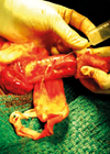
Case 1
A 29-year-old male was attacked and kicked in his left flank. He presented to the emergency department with left flank pain and frank haematuria. He remained haemodynamically stable.
1. What is the suspected diagnosis and what is the investigation of choice?
2. What are the indications for imaging?
3. How are these injuries classified?
4. What grade of injury do you estimate this patient sustained?
Case 2
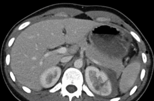
You have been called into the gynaecology operating theatre. They are performing a laparoscopic hysterectomy and suspect the right ureter has been injured.
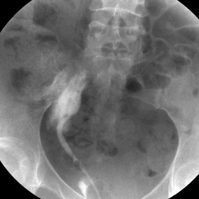
1. What investigation is shown and what is the diagnosis?
2. What is the classification system used for ureteric injury?
3. What are the principles of management?
Case 3
A 35-year-old male had a road traffic accident. He sustained a pelvic fracture with concomitant intra-abdominal visceral injuries, which were stabilised in the operating theatre.
He had blood at his urethral meatus.
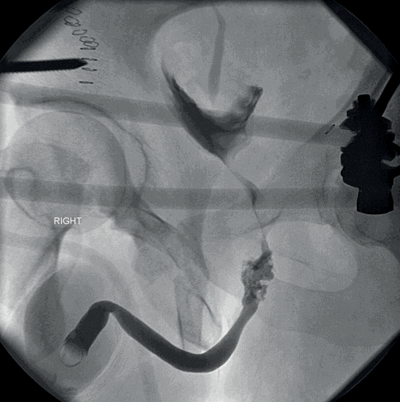
1. What is the investigation shown ?
2. How do you perform this test?
3. What do these images show and how would you manage this patient?
4. What is the AAST classification of urethral injuries?
Urological trauma – answers
Case 1
1. Renal trauma. CT scan with intravenous contrast and delayed excretory imaging.
2. Blunt trauma with visible haematuria (visible or non-visible in paediatric patients). Blunt trauma with non-visible haematuria with hypotension, or the presence of major associated injuries. Rapid deceleration injury. Penetrating trauma regardless of haematuria.
3. The American Association for the Surgery of Trauma (AAST) renal trauma severity scale.
Grade 1
Description of injury: Contusion or non-expanding subcapsular haematoma. No laceration.
Grade 2
Description of injury: Non-expanding peri-renal haematoma. Cortical laceration <1cm deep without extravasation
Grade 3
Description of injury: Cortical laceration >1cm deep without urinary extravasation.
Grade 4
Description of injury: Laceration: through corticomedullary junction into collecting system; or Vascular: segmental renal artery or vein injury with contained haematoma, or partial vessel laceration, or vessel thrombosis.
Grade 5
Description of injury: Laceration: shattered kidney; or Vascular: renal pedicle or avulsion.
Case 2
1. Retrograde ureteropyelogram. Contrast extravasation, demonstrating an iatrogenic injury to the right lower ureter.
2. AAST ureteric injury severity scale.
Grade 1
Type of injury Haematoma
Description of injury Contusion or haematoma without devascularisation
Grade 2
Type of injury Laceration
Description of injury <50% transection
Grade 3
Type of injury Laceration
Description of injury ≥50% transection
Grade 4
Type of injury Laceration
Description of injury Complete transection with <2cm devascularisation
Grade 5
Type of injury Laceration
Description of injury Avulsion with >2cm of devascularisation
3. Surgical principles are: debride necrotic tissue, spatulate ureteral ends, water-tight mucosa-to-mucosa anastomosis with absorbable sutures, internal stenting and external draining. Grade 1 – Conservative +/- ureteric stent. Grade 2/3 – Ureteric stent +/- primary closure with suture. Grade 4/5 – Ureteric reconstruction.
Case 3
1. Retrograde urethrogram.
2. Patient in 30° oblique position. If not possible, supine with AP views acceptable (may miss bulbar urethral extravasation). A 12F catheter is placed in the fossa navicularis and a seal formed with 2mls into the catheter balloon. 20-30mls of water soluble contrast injected slowly via the catheter. Fluoroscopic screening.
3. Partial (incomplete) posterior urethral injury (AAST Grade 3), with contrast passing into the bladder. Gentle attempt at urethral catheterisation can be attempted; if it fails a supra-pubic catheter can be inserted.
4.
Grade 1
Type of injury Contusion
Description of injury Blood at urethral meatus; retrography normal
Grade 2
Type of injury Stretch injury
Description of injury Elongation of urethra without extravasation on urethrography
Grade 3
Type of injury Partial disruption
Description of injury Extravasation of urethrography contrast at injury site with visualisation in the bladder
Grade 4
Type of injury Complete disruption
Description of injury Extravasation of urethrography contrast at injury site with visualisation in the bladder; <2cm of urethra separation
Grade 5
Type of injury Complete disruption
Description of injury Complete transection with ≥2cm urethral separation, or extension into the prostate or vagina