In this series of articles, I am going to show you some of the exhibits contained in the Museum of Urology, hosted on the BAUS website (www.baus.org.uk) this time helped by Hamza, one of our Leicester registrars who I recently remembered reads French!
Mrs A is a 60-year-old, well-nourished lady; for the past year she has been feeling generally tired with right lumbar pain and some urinary frequency. There is a tender palpable swelling, in the right lumbar region and her urine contains pus and evidence of tuberculosis bacilli. What is your diagnosis? Renal TB of the right kidney? So, what are you going to do about it? . . . Oh, just so you know, it’s 1904; there are no CT scanners or renograms and no antibiotics. Your only options are nephrectomy or palliation.
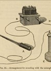
This was the situation Mr Francis Michell Caird (1853–1926), surgeon at the Edinburgh Royal Infirmary, found himself in. Should he remove this lady’s right kidney? What if he was wrong? Caird decided to employ a relatively new instrument from France, Dr George Luys’ urine separator (Figure 1).

Figure 1: Luys’ Urine Separator.
As you can see it looks a little like a urethral sound with a pronounced, sickle-like (Benique) curve. Through the centre runs a chain, which, when the handle is turned, lifts up into that curve. A tubular rubber cover is passed over the end, so when the chain is lifted, it forms a diaphragm which divides the bladder into two. This allows urine from each individual ureter to drain down channels on the right and left of the instrument into test tubes. The urine from each kidney can then be separately examined, giving you more information about their pathology. Mrs A was given an injection of Indigo Carmine to help estimate her differential renal function (Figure 2-I).

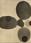
Figure 2-I: Mrs A’s urine results showing a reduced urine output and poorer concentrating ability of the diseased right kidney.
Figure 2-II: Mr WI’s urine showing the suspect right kidney was actually the better one, although neither concentrated well (Both from Caird FM, Trans Med Chir Soc Ed 1905;24:236–45).
As you can see, the right kidney produced less urine (10ml vs. 21ml) and was not able to concentrate the indigo carmine as well. Also, TB bacilli were only found in the right kidney sample. Her right kidney was removed, the specimen showed typical caseating lesions and Mrs A made a good recovery.
As we all know, in the majority of times, a diagnosis is gained from the history and examination; so, you may say to Mr Caird, “I could have told you that without the urine separator, you could have just got on with your nephrectomy”. Which is probably why he went on to write about Mr WI, a 31-year-old, spare man, who also had right renal angle pain and urinary frequency. On examination, he had a tender, palpable and seemingly enlarged right kidney. His right epididymis and the right side of his prostate felt nodular. Another clear case of right renal TB! His separated urine (Figure 2-II) however, showed that the right kidney excreted more urine (29ml vs. 14ml) and slightly more indigo carmine and was thus the better of the two, although both kidneys functioned poorly. Neither kidney showed microscopic evidence of TB. Thankfully, a right nephrectomy (based on history and examination) was not performed, which, as Mr Cains said, would have been a “surgical calamity”.
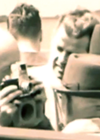
Luys was not the only surgeon to introduce a urine separator. The first was probably that designed by Dr E Lambotte writing in the Brussels Journal of Medicine, Surgery and Pharmacology in 1890. In a series of papers on renal surgery, he noted the importance of knowing the function of the kidneys before surgery, particularly nephrectomy. His urine separator consisted of a double tunnelled tube, at the end of which was a rod and two leaf springs. As the rod was retracted, the two springs spread out forming a circle. The end was enclosed in a child’s rubber balloon, tied on with silk, which formed a diaphragm. When gently pulled back to the bladder neck, urine from the right and left ureters drained down the corresponding tubes for collection. This early attempt was soon abandoned even by Lambotte himself, although he redesigned it in 1903 making it more like Luys’s. Alfred Egon Neumann from Guben, Germany developed one in 1897; it was only suitable for use in women. By 1900, Neumann’s separator had gone through several improvements. The separation was achieved with a bar inserted into the vagina which pushed up into the bladder forming a ridge or fold between the two ureters. The urine separator designed by Malcolm LaSalle Harris (1862–1936) of Chicago, separated the bladder by pushing up a fold from below using a bar inserted into the rectum and hence could be used in men as well as women (Figure 3).

Figure 3: Harris urine segregator showing the uncomfortable looking rectal bar! Image by kind permission of the AUA William P. Didusch Center for Urologic History.
Unsurprisingly perhaps, in a 1907 review on separators, this was said to cause “much pain to the patient” as well as being untrustworthy. Although Ramon Guiteras (1858–1907), a well-respected New York surgeon and American urology pioneer, had “used this instrument a number of times and have never had it fail but once”, he still acknowledged that it could give the wrong results if not accurately placed, leading to “leaking over the barrier”. Harris himself argued that his segregator was superior to those of Neumann, Cathelin’s and Luys’ as it was “very difficult to form a water-tight joint by pressing a smooth edge against a mucous membrane naturally thrown into folds”.
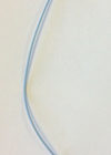
George Luys (1870–1953) of the Laboisière Hospital, Paris, introduced his separator in 1901 and Fernand Cathelin (1873–1960), also from France, devised his ‘diviseur vésical’ in 1902. Looking similar to a curved sound it was hollow and contained a folded India-rubber leaf held open by a wire loop (Figure 4). When the loop was launched out of the instrument it formed a septum dividing the bladder.

Figure 4: Cathelin’s diviseur vésical, the wire loop is extended, but the rubber diaphragm it used to hold is long gone. Image by kind permission of the EAU History Office.
Historically, I suspect urine separators were short lived. At around the same time cystoscopy was developing, and this was soon followed by ureteric catheterisation under vision. Cannulation of individual ureters being greatly helped by the invention of the Albarran lever. However, the skill required to cystoscope someone and cannulate both ureters was greater than merely passing a urine separator and the kit was more expensive. Any decent surgeon could pass a silver catheter or sound, but it needed a specialist, a urologist, to master these early cystoscopes. Nevertheless, direct ureteric cannulation for individual renal sampling soon overtook the blind separators. Also, radiology was progressing and X-rays, which could show a calcified tuberculous kidney, retrograde studies and intravenous urograms were not long behind.