1. What does LASER stand for?
2. What elements are involved and how is a LASER created?
3. The following cases involve laser surgery. Can you diagnose the pathology, identify which type of lasers can be used, and their wavelengths?
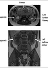
a) A 65-year-old male presents to clinic with worsening LUTS despite being on dual medical therapy. His MRI prostate shows 105cc prostate with no concerning lesions. He would like to consider surgical intervention. See Figure 1.
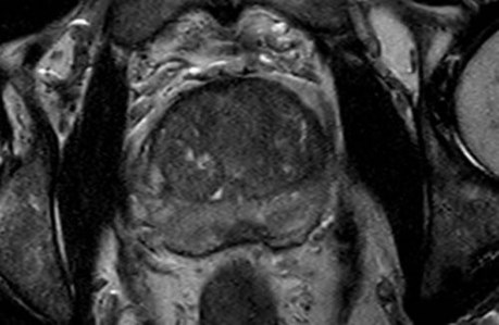
Figure 1.
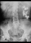
b) A 35-year-old male presents with episodes of recurrent urinary tract infection. A CTKUB is conducted and pathology identified, for which he would prefer endoscopic intervention. See Figure 2.
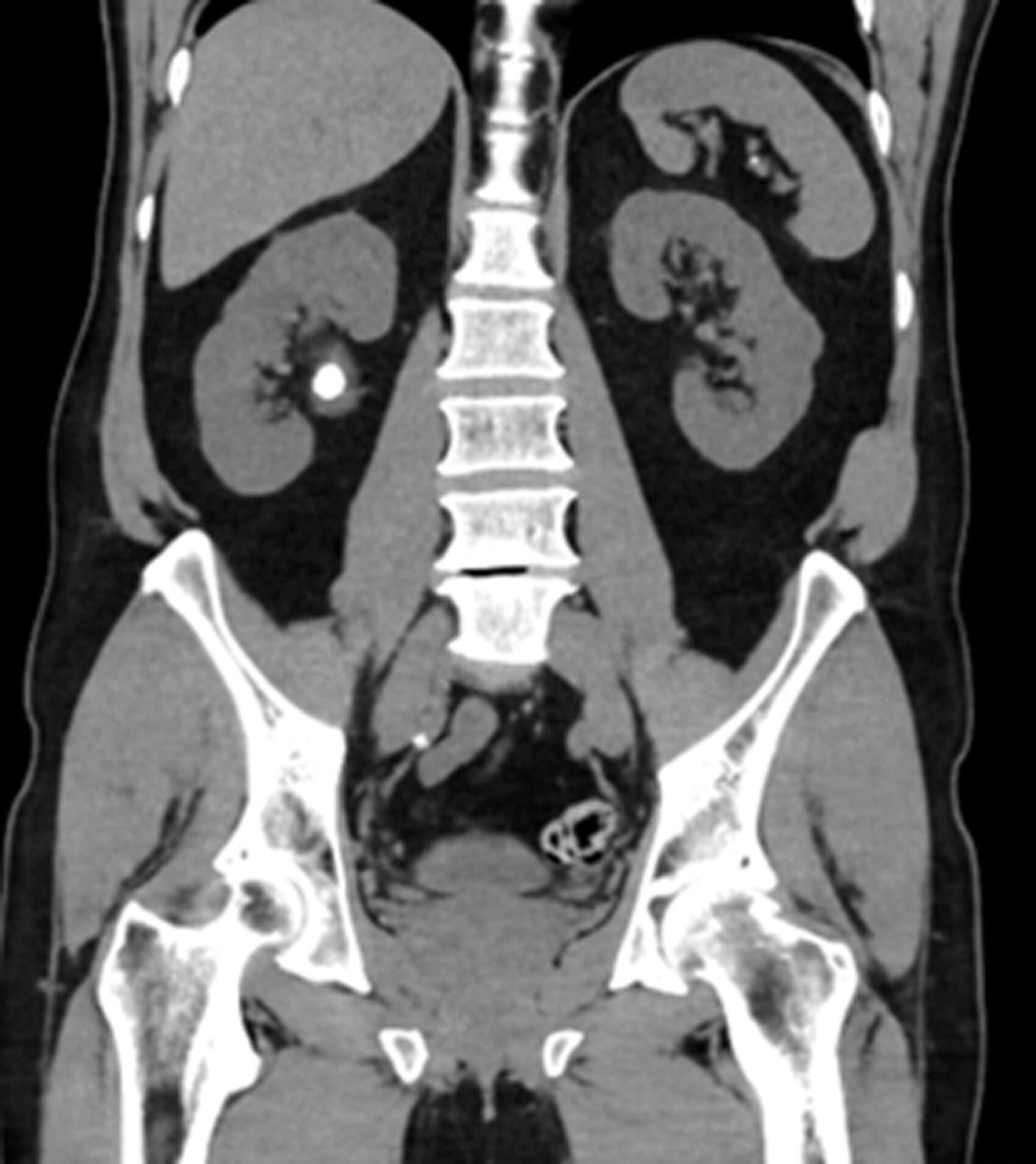
Figure 2.
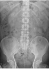
c) An 80-year-old male with significant comorbidities (AF, IHD, HTN, COPD, frailty) presents with haematuria. CT urogram has been performed. Given his high risk for general anaesthesia, what treatment modality using laser could be considered? See Figure 3.
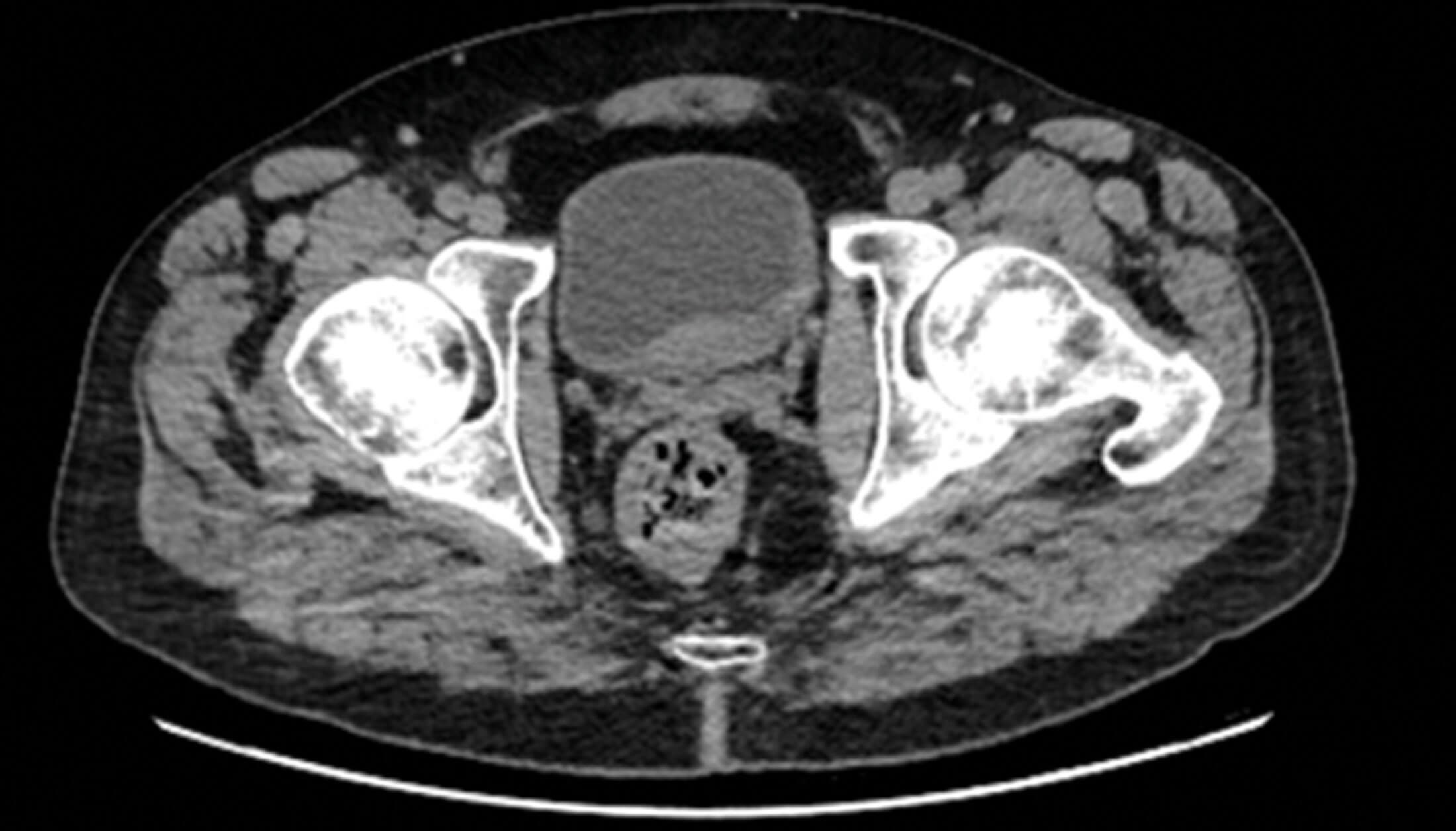
Figure 3.
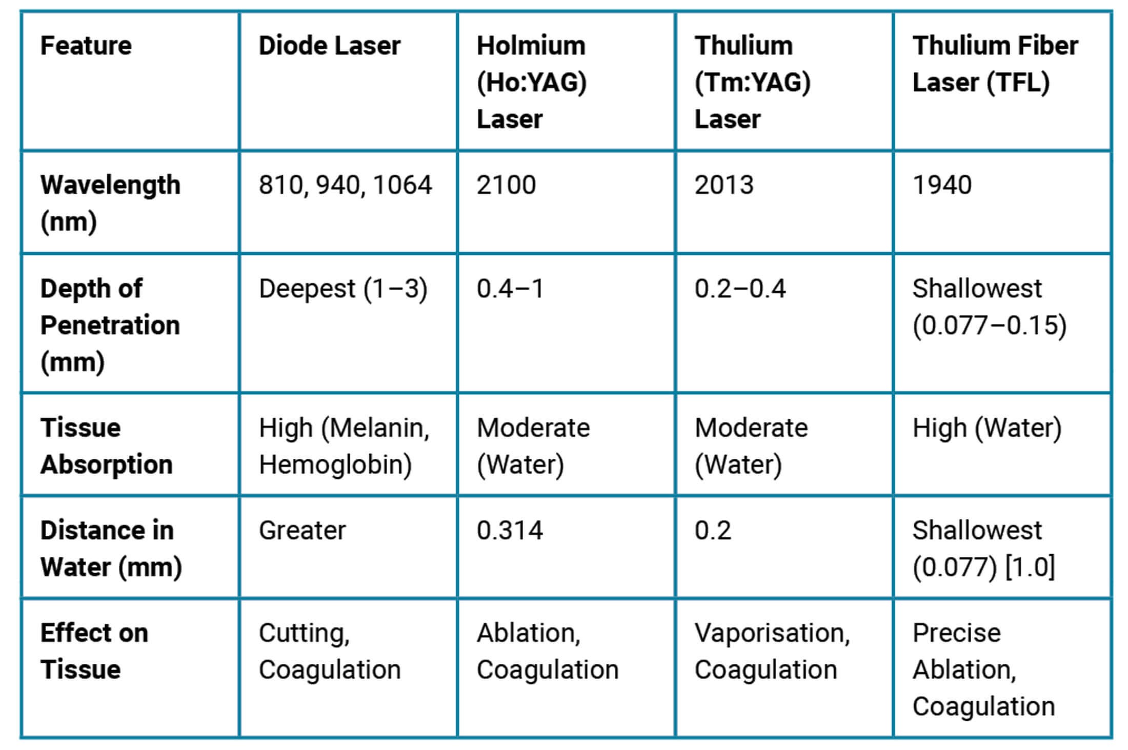
4. Can you arrange the following Lasers (Holmium, Thulium, Thulium fibre, Diode) based on the depth of tissue penetration, tissue absorption, distance in water and effect on tissue?
Answers
1. Light Amplification by Stimulated Emission of Radiation.
2. The elements of a laser include a light source, gain medium such as Nd:YAG crystal containing Holmium ions and reflective mirrors [1]. A laser is created by:
- A chamber containing a gain or amplification medium (e.g. Holimium: Yag) is enclosed by a complete mirror on one end, and a mirror with a pinhole in the middle at another end.
- An energy source is used to supply energy to the gain medium.
- The gain medium consists of numerous atoms. When the energy is pumped into the medium from the energy source, electrons in the atoms move to a higher orbit by absorption of energy. When these electrons come back to their ground state, they emit a photon of light. This photon then keeps bouncing on and off the medium due to reflection from the mirrors, releasing more photons, which eventually escape through the pinhole to form a concentrated laser beam.
3. a) This patient has Benign Prostatic Hyperplasia and could undergo the following [2]:
- ThuLEP: Thulium Laser Enucleation of the Prostate (2013nm)
- ThuFLEP: Thulium Fiber Laser Enucleation of the Prostate (1940nm)
- HoLEP: Holmium Laser Enucleation of the Prostate (2100nm)
- GLL PVP: Green Light Laser Photo Selective vaporization of Prostate (532nm).
b) This patient has a right renal pelvic stone, and can undergo ureteroscopic laser stone fragmentation using a Holmium or Thulium laser.
c) He has a bladder tumour, likely to be Transitional cell carcinoma. He could undergo Transurethral Laser Ablation (TULA) of the tumour under LA, using diode laser (980nm or 1470nm) [3].
4.
References
1. Urology Resources. FRCS Notes – lasers in urology [video] (June 2018).
www.youtube.com/watch?v=Mvuyu_tLVC8
2. Son H, Song SH, Paick JS. Current laser treatments for benign prostatic hyperplasia. Korean J Urol 2010;51(11):737–44.
3. Hameed F, Anwaar A. Bladder tumor ablation with 980-nm and 980-/1470-nm diode lasers: a retrospective study. Afr J Urol 2024;30(1).
https://doi.org/10.1186/s12301-023-00404-z
4. Zarrabi A, Gross AJ. The evolution of lasers in urology. Ther Adv Urol 2011;3(2):81–9.
[All links last accessed May 2026].