- Click for Part 1 and Part 2 on this topic -
Case scenario
A 36-year-old man with a known renal stone attends your stone clinic following a surveillance CT KUB with worsening intermittent right flank pain.

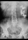
Figures 1 and 2.
1. Please describe the findings in Figure 1 and Figure 2.
2. What management options are available to this patient, and which anatomical factors should be considered when counselling him on his options?

Figure 3 and 4.
3. Following counselling the patient opts for ureteroscopy and laser lithotripsy to manage his stone. What equipment is shown in Figure 3 and Figure 4?
4. How does flexible and navigable suction (FANS) assisted flexible ureteroscopy work and what proposed benefits might this technology confer?
Answers
1. Figure 1 is an axial image of an unenhanced, low dose CT KUB in the soft tissue window, demonstrating an 11mm non obstructing calculus in the right lower renal pole. Figure 2 shows the corresponding coronal reconstruction confirming the lower pole stone position and demonstrates a hypoplastic left kidney.
2. Given the size of the calculus along with worsening symptoms, it is reasonable to recommend active treatment preferentially to observation. EAU guidelines for lower pole renal stones (see image below) suggest all active management options can be considered but recommend taking certain anatomical factors affecting stone clearance rates into consideration when deciding between ESWL Vs URS/PCNL [1].

Figures 5: EAU guidelines for lower pole renal stones.
These anatomical factors include the infudibulo-pelvic angle (IPA), width and length and the ratio between the two, all of which have a cumulatively negative effect on the stone clearance rate after ESWL when deemed ‘unfavourable’ [2,3]. Whilst no exact values have been set as guidance, IPA <90 degrees with infundibular length and width >3cm and <5mm respectively are associated with reduced clearance rates following ESWL [4].
3. Figure 3 is of a flexible and navigable suction (FANS) access sheath and Figure 4 is a close-up of the y-connector which enables continuous suction alongside ureteroscopic access.
4. A FANS access sheath incorporates a flexible and bendable tip allowing for greater manoeuvrability and improved navigation during flexible ureteroscopy, improving access to complex renal calyces compared to traditional access sheaths [4]. Additionally, connecting a suction vacuum device can assist with fragment evacuation and maintain lower intrarenal pressures during surgery. Evidence suggests that using FANS access sheaths may achieve superior stone free rates following ureteroscopy and laser lithotripsy along with a decreased necessity for stone basket utilisation and a reduction in post-operative fever rates [5].
References
1. Skolarikos A, Davis NF, Gambaro G, et al. EAU Guidelines on Urolithiasis.2026.
https://uroweb.org/guidelines/urolithiasis/chapter/guidelines
[Link last accessed April 2026]
2. Sumino Y, Mimata H, Tasaki Y, et al. Predictors of lower pole renal stone clearance after extracorporeal shock wave lithotripsy. J Urol 2002;168(4 Pt 1):1344–7.
3. Elbahnasy A, Shalhav A, Hoenig D, et al. Lower caliceal stone clearance after shock wave lithotripsy or ureteroscopy: the impact of lower pole radiographic anatomy. J Urol 1998;159(3):676–82.
4. Zhu W, Liu S, Cao J, et al. Tip bendable suction ureteral access sheath versus traditional sheath in retrograde intrarenal stone surgery: an international multicentre, randomized, parallel group, superiority study. EClinicalMedicine 2024;74:102724.
5. Alnadhari I, Abdeljaleel O, Ali O, et al. Comparison between flexible and navigable suction ureteral access sheath and standard ureteral access sheath during flexible ureteroscopy for the management of kidney stone: systematic review and meta-analysis. BMC Urol 2025;25(1):115.
Declaration of competing interests: None declared.