Features
Seminal vesicle calculi
Epidemiology Seminal vesicle calculi are uncommon with just over 100 cases being reported in the literature, although the true incidence is likely to be higher [1-9]. Patients usually present aged between 30 and 45 years old and although the pathogenesis...

The emerging role of physician associates in urology
The physician associate (PA) is a new role in the NHS which has expanded across medical and surgical specialties to include urology. In the USA, it has long been an established field of practice where physician assistants work autonomously within...
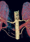
Radiological appearances of renal vascular anatomical variants
The purpose of this article is to explain and illustrate common renal vascular variants that can be depicted with imaging. Renal vessels commonly present a wide range of variations [1]; before major renal or vascular surgery is undertaken, accurate portrayal...
Radiological appearances of non-vascular renal anatomical variants
Anatomical variants of the renal tract are common and, although often asymptomatic, may present with complications. It is essential to identify anatomical variants, as this may have an impact upon surgical planning and management. This article aims to demonstrate radiological...
Reflections on 20 years as an Army Reserve doctor: live a life less ordinary
It seems a very short time ago that my predecessor recruited me into my regiment as a surgical senior house officer during a varicose vein operation in a cottage hospital in Stroud, informing me that I would be only the...
Guide to gaining approval for a clinical study
This article focuses on gaining approval for clinical research involving NHS patients, although the principles can be applied to other types of research. It can be quite a daunting process for the uninitiated applicant. Often it can be made less...

What to expect when meeting a statistician
There are a growing number of statisticians working closely with medics from all specialties. They have different training but they are driven by the same goal: to perform high quality evidence-based clinical research [1,2]. In a perfect world we would...
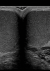
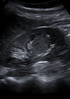
Testicular microlithiasis
Introduction Testicular microlithiasis (TML) was originally described in 1970 in a healthy four-year-old boy [1] and the first paper regarding microlithiasis as an entity seen on ultrasound was published in 1987 [2]. Testicular microlithiasis is seen on ultrasound as small,...
An MA in medical education – is it for you?
I have recently completed a three-year MA in medical education at the University of Winchester, which has been an edifying experience. The following article may appeal to readers who are considering such a venture. I have been a consultant for...
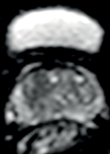
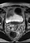
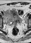
Use of MRI in the evaluation of prostate cancer: part 2
Diffusion weighted imaging and contrast enhanced imaging Introduction Magnetic resonance imaging (MRI) is widely used in localisation, staging and post-treatment follow-up of prostate cancer. In the previous issue, we discussed the usefulness of MRI in depicting prostate anatomy and pairing...
Use of MRI in the evaluation of prostate cancer: part 1
Introduction Prostate cancer remains the most commonly diagnosed cancer in males and the second leading cause of cancer related deaths in UK men, after lung cancer [1]. The incidence of prostate cancer in the UK has shown a rapid increase...
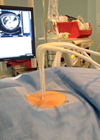
Image-guided renal cryoablation
Introduction There has undoubtedly been a dramatic increase in the number of patients diagnosed with small renal masses in recent years [1]. The rapidly expanding use of CT has led to a large number of incidental diagnoses, but increasing longevity...

